|
※サムネイル画像をクリックすると拡大画像が表示されます。
Fig.1. Testeis specific expression of Calreticulon-3 as examined in various tissues by western blotting with anti-CALR3 antibody.
The various tissues were excised and homogenized in lysis buffer containing 1% TritonX100 and then placed on ice for 1 h.
These extracts were centrifuged, and the supernatants were collected and analyzed by western blotting with anti-CALR3 antibody
at 1/1,000 dilution.
1. Brain. 2. Lung. 3. Heart. 4. Thymus. 5. Liver. 6. Spleen. 7. Kidney. 8. Muscle. 9. Ovary. 10. Testis. 11. Sperm
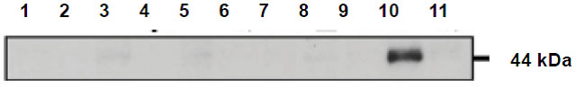
Fig.2. Identification of Calreticulon-3 protein by western blotting with anti-CALR3 antibody.
Embryonic fibroblast cells prepared from Calr3 -/- mouse were transfected with a plasmid expressing Calr3. The cell lysate was analyzed by western blotting with anti-CALR3 antibody at 1/1,000 dilution.
1. Mock-infected cell lysate as a negative control.
2. Cell lysate transfected with a plasmid expressing Calr3.
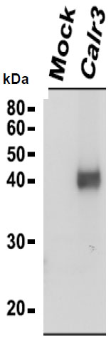
Fig.3 Immunoprecipitation of Calreticulon-3 protein with anti-CALR3 antibody.
Lysates of wild-type mouse testis were immunoprecipitated with anti-CALR3 antibody and the precipitates were analyzed by western blotting with the same antibody.
1. Input testis lysate
2. Precipitated with preimmune serum
3. Precipitated with anti-CALR3 antibody
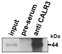
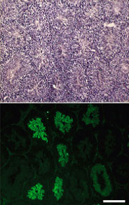
Fig.4 Immunofluorescence staining of a testicular section with anti-CALR3 antibody.Sequential sections were stained with hematoxylin and eosin (upper panel). CALR3 was detected in elongating spermatids (lower panel). Testis was collected from adult mouse and fixed in 4% paraformaldehyde/PBS overnight at 4 °C, cryopreserved in graded 10?30% sucrose, and embedded. Frozen sections (8 μm) were mounted on aminopropyl triethoxysilane-coated glass slides. Primary antibody was used at 1/100 and as secondary antibody, Alexa Fluor 488 conjugated goat anti-rabbit IgG was used. Scale bar is 200 μm.
|

