|
※サムネイル画像をクリックすると拡大画像が表示されます。
Fig.1. Detection of SUMO-2/3 by Western blotting with anti-SUMO2/3 antibody (3H12).
High molecular multiple bands were observed in HeLa total cell extract.
As secondary antibody, Alkaline phosphatase conjugated anti-rat IgG was used.
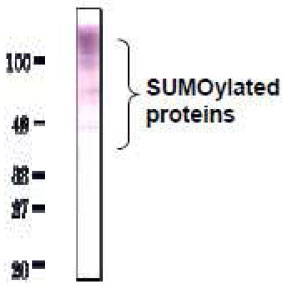
Fig.2. Detection of SUMO-2/3 in whole cell extracts of mammalian cells by Western blotting with anti-SUMO2/3 antibody (3H12).
1. MCF-7 (human breast cancer cell line)
2. NIH3T3 (mouse fibroblast cell line)
3. CHO (Chinese Hamster Ovary cell)
10-20% gradient gel was used for SDS-PAGE. Wet blotting method was employed. Anti-SUMO-2/3 antibody (3H12) was used at 1/1,000 dilution. As a second antibody, goat anti-rat IgG antibody conjugated with HRP was used at 5,000 dilution.
*Arrow indicates unconjugated SUMO-2/3 proteins. SUMO-2/3 proteins conjugate numerous proteins in vivo, and SUMOylation states vary depending on the kinds of cells and physiological states of them.

Fig.3 Immunofluorescence staining of SUMO-2/3 with the anti-SUMO2/3 antibody (3H12) in the mouse primary neural progenitor cells. DNA was stained with Hoechst.

Fig.4 SUMO-2/3 foci detection in C-33A cells by imunofluorescence staining with anti-SUMO-2/3 antibody (3H12).
Cells were fixed with 4% paraformaldehyde and permeabilized with 0.25 TritonX-100. As secondary antibody, Alexa 488 conjugated donkey anti-rat IgG was used. Cells were analyzed using Olympus IX71 microscope and Lumina Vision software (Mitani Co., Tokyo)

Fig.5 Immunohistochemistry of Coronal section of E16.mouse cerebral cortex.
Coronal section was immunostained with anti-SUMO-2/3 antibody (3H12). DNA was stained with Hoechst.

|

