|
※サムネイル画像をクリックすると拡大画像が表示されます。
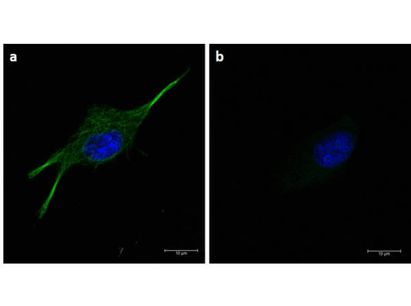
Immunofluorescence microscopy of α-tubulin in U-87 MG cells using FITC-conjugated Fluorescent TrueBlot(R) anti-mouse IgG (p/n 18-0217-32) for detection. U87-MG cells were fixed with 100% methanol, blocked (5% rat serum/0.3% Triton X-100) for 1hr, then incubated with 15μg/mL of anti-alpha-tubulin primary antibody (p/n 200-301-880) at 4°C overnight. Following 3 washes in 1X PBS for 5min each, 5μg/mL of Fluorescent TrueBlot(R) anti-mouse IgG Fluorescein was added and allowed to incubate for 1hr at room temperature. 5μg/mL of Fluorescent TrueBlot(R) anti-mouse IgG FITC was added and allowed to incubate for 1hr at room temperature. Nucleus was counterstained with DAPI present in mounting medium. The predicted main localization is microtubules. Image taken at 63X magnification. (a) Merged α-tubulin (green)/DAPI (blue) image shown (b) secondary only.
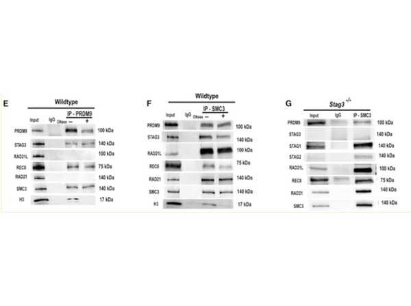
(E&F) Co-immunoprecipitation of PRDM9 with chromosomal axis proteins STAG3, REC8, and SMC3 from wildtype 14-dpp spermatocytes (E), confirmed by reverse co-immunoprecipitation with anti-SMC3 antibody (F). Input extract, 20 μg (2.5 μg in the RAD21,SMC3 and H3 blots); lane 2, 20 μg extract coIP with nonimmune IgG; αPRDM9 and αSMC3 (F), 20 μg extract coIP with the respective antibody, either not-treated ("-") or treated ("+") with DNase. Histone H3 IP was used to detect efficiency of DNase treatment. The antibodies used to detect specific proteins are shown on the left and the relevant molecular weights are shown on right of each blot. For quantified values of protein bands please refer to?Table S1?and?S2. (G) Co-immunoprecipitation of PRDM9 and cohesin proteins from?Stag3-/-?spermatocytes. This analysis confirmed that PRDM9 can be associated with other cohesin subunits in absence of STAG3, and demonstrates the protein-protein interaction of REC8 and RAD21L with other cohesin subunits in absence of STAG3 previously reported [7]. Input extract, 20 μg (2.5 μg in the STAG1, STAG2, RAD21 and SMC3 blots); lane 2, 20 μg extract coIP with nonimmune IgG and lane 3, 20 μg extract coIP with SMC3 antibody. The relevant molecular weights are shown on right of each blot. Fig 1. PMID:?30853435
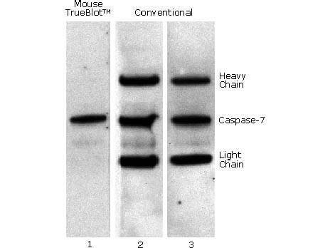
Mouse TrueBlot(R) IP / Western Blot: Caspase 7 was immunoprecipitated from 0.5 ml of 1x10e7 Jurkat cells/ml with 5 ug mouse anti-human Caspase 7. Precipitate from 1x10e6 cells was subjected to electrophoresis, transferred to an PVDF membrane, and Western blotted with anti-Caspase 7 using Mouse TrueBlot(R) ULTRA: Anti-Mouse Ig HRP (Lane 1) or conventional HRP-conjugated anti-mouse antibody (Lane 2) - note the detection of the heavy and light chains of the immunoprecipitating antibody in Lane 2 but not in Lane 1. When Lane 1 is re-immunoblotted using conventional HRP-conjugated anti-mouse polyclonal antibody (Lane 3), the heavy and light chains are now detected, confirming that although the immunoprecipitating heavy and light chains are present, Mouse TrueBlot(R) ULTRA: Anti-Mouse Ig HRP detects only native antibody and not denatured heavy and light chains.
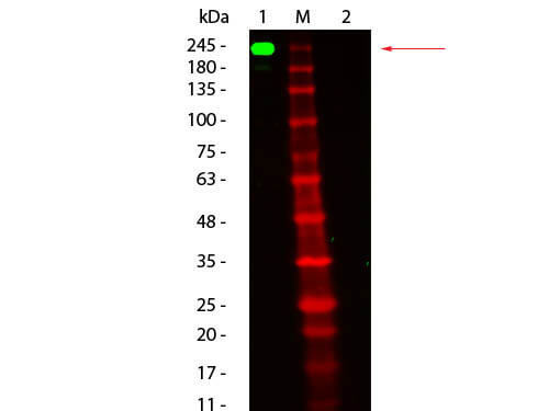
Western Blot of Fluorescent TrueBlot(R): Anti-Mouse Ig Fluorescein. Lane 1: Mouse IgG, Non-reduced. Lane 2: Mouse IgG, Reduced. Load: 50 ng per lane. Primary antibody: none. Secondary antibody: Fluorescent TrueBlot(R): Anti-Mouse Ig Fluorescein at 1:1,000 for 60 min at RT. Block: MB-070 for 30 min at RT. Predicted/Observed size: 160 kDa for Mouse IgG, Non-reduced. Migrates at slightly higher molecular weight. Other band(s): none.
|
|
|
|
Immunofluorescence microscopy of α-tubulin in U-87 MG cells using FITC-conjugated Fluorescent TrueBlot(R) anti-mouse IgG (p/n 18-0217-32) for detection. U87-MG cells were fixed with 100% methanol, blocked (5% rat serum/0.3% Triton X-100) for 1hr, then incubated with 15μg/mL of anti-alpha-tubulin primary antibody (p/n 200-301-880) at 4°C overnight. Following 3 washes in 1X PBS for 5min each, 5μg/mL of Fluorescent TrueBlot(R) anti-mouse IgG Fluorescein was added and allowed to incubate for 1hr at room temperature. 5μg/mL of Fluorescent TrueBlot(R) anti-mouse IgG FITC was added and allowed to incubate for 1hr at room temperature. Nucleus was counterstained with DAPI present in mounting medium. The predicted main localization is microtubules. Image taken at 63X magnification. (a) Merged α-tubulin (green)/DAPI (blue) image shown (b) secondary only.
|
|
| 別品名 |
Anti-Mouse IgG FITC, TrueBlot, FITC TrueBlot ULTRA, Fluorescein TrueBlot, TrueBlot for IP/WB, TrueBlot for immunoprecipitation, TrueBlot for western blotting, Fluorescent TrueBlot, Ms TrueBlot
|
| 交差種 |
Mouse
|
| 適用 |
Western Blot
Immuno Fluorescence
Immunoprecipitation
|
| 免疫動物 |
Rat
|
| クローン |
eB144
|
| 標識物 |
Fluorescein Isothiocyanate
|
| 精製度 |
Affinity Purified
|
| 純度 |
Ig-PG
|
| 参考文献 |
[[Cosmo Reference Collection]][Pub Med ID]30853435
|
| [注意事項] |
濃度はロットによって異なる可能性があります。メーカーDS及びCoAからご確認ください。
|
|
| メーカー |
品番 |
包装 |
|
RKL
|
18-0217-32
|
100 UL
|
※表示価格について
| 当社在庫 |
なし
|
| 納期目安 |
約10日
|
| 法規制 |
毒
|
| 保存温度 |
4℃
|
|
※当社では商品情報の適切な管理に努めておりますが、表示される法規制情報は最新でない可能性があります。
また法規制情報の表示が無いものは、必ずしも法規制に非該当であることを示すものではありません。
商品のお届け前に最新の製品法規制情報をお求めの際はこちらへお問い合わせください。
|
※当社取り扱いの試薬・機器製品および受託サービス・創薬支援サービス(納品物、解析データ等)は、研究用としてのみ販売しております。
人や動物の医療用・臨床診断用・食品用としては、使用しないように、十分ご注意ください。
法規制欄に体外診断用医薬品と記載のものは除きます。
|
|
※リンク先での文献等のダウンロードに際しましては、掲載元の規約遵守をお願いします。
|
|
※CAS Registry Numbers have not been verified by CAS and may be inaccurate.
|


