|
※サムネイル画像をクリックすると拡大画像が表示されます。
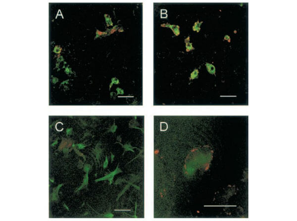
Immunofluorescence of Rabbit Anti-TACE Antibody. Cells: Rat Cortical Neurons. (A) Control cultures show TACE immunoreactivity at the cellular membrane of some microglial cells. (B) Glutamate-exposed cultures show that most microglial cells express TACE immunoreactivity. (C) Control cultures show that TACE immunostaining does not colocalize with astrocytes [glial fibrillary acidic protein (GFAP)-positive cells]. (D) Astrocyte (GFAP-positive cell) showing TACE immunoreactivity in its surface after treatment with glutamate. Double immunostaining of control and glutamate-exposed rat cortical cultures. (Hurtado et al., 2002).
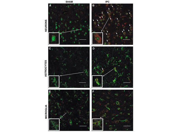
Immunofluorescence of Rabbit Anti-TACE Antibody. Cells: Rat Brain. Cellular localization of TACE. Double immunofluorescence staining of brain sections from sham-operated (SHAM; A, C, E) and IPC-exposed animals (IPC; B, D, F) of TACE (red) and the cellular markers (green) NeuN (neurons; A, B), GFAP (astrocytes; C, D) and L. esculentum lectin (microglia and endothelium; E, F). White arrows indicate TACE-positive cells. (Pradillo et al, 2005).
Immunofluorescence of Anti-TACE Antibody. Cells: HeLa Cells. Primary Antibody: Anti-TACE 10μg/mL. Secondary Antibody: Goat Anti-Rabbit IgG 1:500. Fixation: 4% paraformaldehyde.
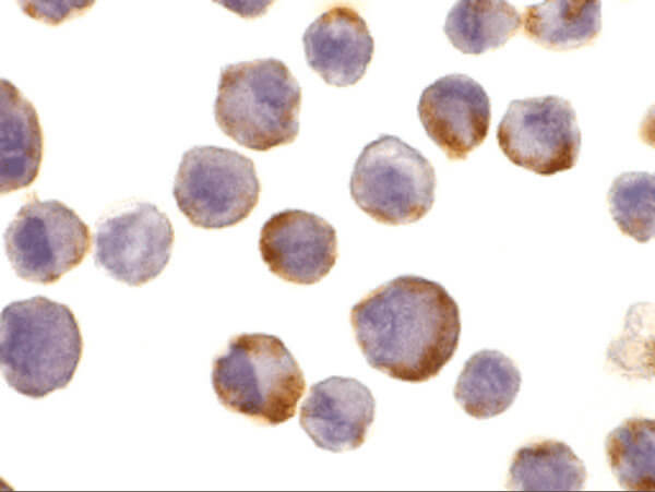
Immunocytochemistry of Rabbit Anti-TACE Antibody. Cells: HeLa cells. Primary Antibody: Anti-TACE antibody at 10 μg/ml overnight at 2-8°C. Fixation: formaldehyde and blocked with 10% serum for 1 h at RT. Antigen Retrieval: heat mediation with a citrate buffer (pH6). Secondary Antibody: goat anti-rabbit IgG H&L (HRP) at 1:250. Counter stained with Hematoxylin.
Western Blot of Rabbit Anti-TACE Antibody. Lane (A, D): HeLa. Lane (B, E): Jurkat. Lane (C, F): Raji. Lanes A-C are in the absence or Lanes D-F are in the presence of blocking peptide. Primary Antibody: Anti-TACE at 1μg/mL RT for 1hr. Secondary Antibody: Goat Anti-Rabbit IgG HRP at 1:10,000. Blocking 5% NFDM/TBST. Expect: ~93kDa.
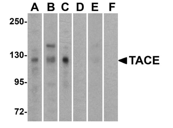
Western Blot of Rabbit Anti-TACE Antibody. Loading: 15 μg of lysates per lane. Primary Antibodies: Anti-TACE 600-401-H24 (0.5 μg/mL), TACE 22001 (2 μg/mL), and GAPDH (0.02 μg/mL), for 1h incubation at RT. Block: 5% NFDM/TBST. Secondary: Goat anti-rabbit IgG HRP conjugate at 1:10,000. Expect: ~93kDa.

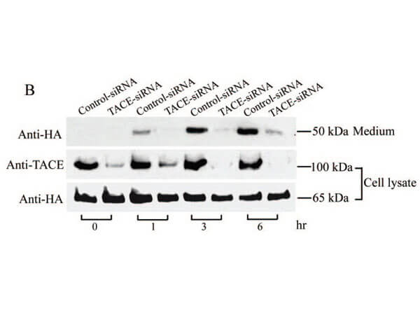
Western Blot of Rabbit Anti-TACE Antibody. Monkey COS cells stably expressing Pref-1A were transfected with control siRNA or TACE siRNA. TACE was detected in lysates by using the anti-TACE antibody. TACE expression levels were markedly reduced in TACE knockdown cell lysate. (Wang et al., 2006).
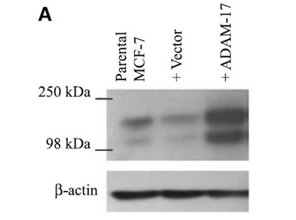
Western Blot of Anti-TACE (ADAM-17) Antibody. ADAM-17 (TACE) protein expression, following transfection of vector and ADAM-17 cDNA, was examined by immunoblot analysis with anti-ADAM-17 antibodies in MCF-7 cells. Increased ADAM-17 was detected in ADAM-17 transfected cells. (McGowan et al., 2007).
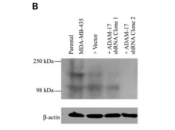
Western Blot of Rabbit Anti-TACE (ADAM-17). ADAM-17 (TACE) protein expression in MDA-MB-435 Cells, following transfection with ADAM-17 shRNA (two clones) or neomycin-resistant negative control vector, was examined by immunoblot analysis with anti-ADAM-17 antibodies. (McGowan et al., 2007).
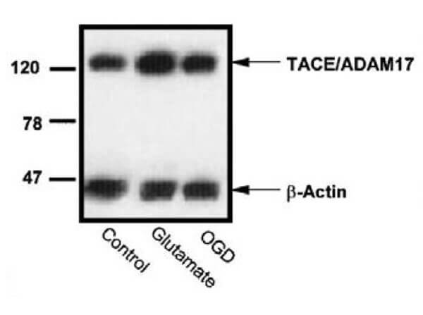
Western Blot of Anti-TACE Antibody. Effect of oxygen?glucose deprivation (OGD) or glutamate on the levels of TACE/ADAM17 in rat cortical cultures. (Hurtado et al., 2002). Western blot analysis of TACE in Rat Cortical Neuron homogenates from control, glutamate, and OGD-exposed cultures from a representative experiment.
|
|
|
|
Immunofluorescence of Rabbit Anti-TACE Antibody. Cells: Rat Cortical Neurons. (A) Control cultures show TACE immunoreactivity at the cellular membrane of some microglial cells. (B) Glutamate-exposed cultures show that most microglial cells express TACE immunoreactivity. (C) Control cultures show that TACE immunostaining does not colocalize with astrocytes [glial fibrillary acidic protein (GFAP)-positive cells]. (D) Astrocyte (GFAP-positive cell) showing TACE immunoreactivity in its surface after treatment with glutamate. Double immunostaining of control and glutamate-exposed rat cortical cultures. (Hurtado et al., 2002).
|
|
| 別品名 |
ADAM17, ADAM18, CD156B, CSVP, NISBD, NISBD1, TACE
|
| 交差種 |
Human
Rat
|
| 適用 |
Western Blot
Enzyme Linked Immunosorbent Assay
Immunohistochemistry
Immuno Fluorescence
|
| 免疫動物 |
Rabbit
|
| 抗原部位 |
C-terminus
|
| 標識物 |
Unlabeled
|
| 精製度 |
Affinity Purified
|
| GENE ID |
6868
|
| Accession No.(Gene/Protein) |
NP_003174, P78536
|
| Gene Symbol |
ADAM17
|
| 参考文献 |
Black RA, Rauch CT , Kozlosky CJ, et al. A metalloproteinase disintegrin that releases tumour-necrosis factor-α from cells. Nature 1997;385:729-733 Moss ML, Jin SL, Milla ME, et al. Cloning of a disintegrin metalloproteinase that processes precursor tumour-necrosis factor-α. Nature 1997;385:733-736 Mizui Y, Yamazaki K, Sagane K, Tanaka I. cDNA cloning of mouse tumor necrosis factor-α converting enzyme (TACE) and partial analysis of its promoter. Gene 1999;233:67-74 Peschon JJ, Slack JL, Reddy P, et al. An essential role for ectodomain shedding in mammalian development. Science 1998;282:1281-4
|
| [注意事項] |
濃度はロットによって異なる可能性があります。メーカーDS及びCoAからご確認ください。
|
|
| メーカー |
品番 |
包装 |
|
RKL
|
600-401-H24
|
100 UG
|
※表示価格について
| 当社在庫 |
なし
|
| 納期目安 |
約10日
|
| 保存温度 |
-20℃
|
|
※当社では商品情報の適切な管理に努めておりますが、表示される法規制情報は最新でない可能性があります。
また法規制情報の表示が無いものは、必ずしも法規制に非該当であることを示すものではありません。
商品のお届け前に最新の製品法規制情報をお求めの際はこちらへお問い合わせください。
|
※当社取り扱いの試薬・機器製品および受託サービス・創薬支援サービス(納品物、解析データ等)は、研究用としてのみ販売しております。
人や動物の医療用・臨床診断用・食品用としては、使用しないように、十分ご注意ください。
法規制欄に体外診断用医薬品と記載のものは除きます。
|
|
※リンク先での文献等のダウンロードに際しましては、掲載元の規約遵守をお願いします。
|
|
※CAS Registry Numbers have not been verified by CAS and may be inaccurate.
|
