|
※サムネイル画像をクリックすると拡大画像が表示されます。
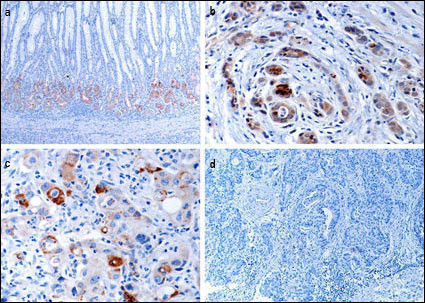
Immunohistochemical of EphB1 in gastric cancer tissues. a EphB1 protein expressed in normal mucosa at the glandular compartment and in a decreasing gradient from the glandular compartment to the foveolar compartment. b EphB1 protein focally positively stained in well-differentiated gastric cancer cells. c EphB1 protein is focally positive in poorly differentiated gastric cancer cells. d Loss of EphB1 expression in gastric cancer cells.(Provided by Jian-dong Wang,Department of Pathology Nanjing Jinling Hospital/Nanjing University School of Medicine)
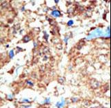
Formalin-fixed and paraffin-embedded human cancer tissue reacted with the primary antibody, which was peroxidase-conjugated to the secondary antibody, followed by DAB staining. This data demonstrates the use of this antibody for immunohistochemistry; clinical relevance has not been evaluated. BC = breast carcinoma; HC = hepatocarcinoma.
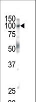
Western blot of lysates from A431, HeLa, MDA-MB-453 cell line (from left to right), using EPHB1 Antibody (H970). Antibody was diluted at 1:1000 at each lane. A goat anti-rabbit IgG H&L (HRP) at 1:5000 dilution was used as the secondary antibody. Lysates at 35ug per lane.

Western blot of anti-EphB1 antibody in mouse brain tissue. EphB1 (arrow) was detected using purified antibody. Secondary HRP-anti-rabbit was used for signal visualization with chemiluminescence.
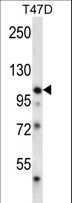
EPHB1 Antibody (H970) western blot of T47D cell line lysates (35 ug/lane). The EPHB1 antibody detected the EPHB1 protein (arrow).
|
|
|
|
Immunohistochemical of EphB1 in gastric cancer tissues. a EphB1 protein expressed in normal mucosa at the glandular compartment and in a decreasing gradient from the glandular compartment to the foveolar compartment. b EphB1 protein focally positively stained in well-differentiated gastric cancer cells. c EphB1 protein is focally positive in poorly differentiated gastric cancer cells. d Loss of EphB1 expression in gastric cancer cells.(Provided by Jian-dong Wang,Department of Pathology Nanjing Jinling Hospital/Nanjing University School of Medicine)
|
|
| 別品名 |
EPHB1, Cek6, EK6, ELK, Ephrin type-B receptor 1, Hek6, EPHT2, NET, EPH receptor B1, EPH tyrosine kinase 2, EPH-like kinase 6, Soluble EPHB1 variant 1
|
| 種由来 |
Human
|
| 交差種 |
Human
Mouse
|
| 適用 |
Western Blot
IHC paraffin embedding section
Immunohistochemistry
|
| 免疫動物 |
Rabbit
|
| 抗原部位 |
a.a.955-984
|
| 標識物 |
Unlabeled
|
| 精製度 |
Ig fraction - Ammonium Sulphate
|
| GENE ID |
2047
|
| Gene Symbol |
EPHB1
|
|
| メーカー |
品番 |
包装 |
|
LSP
|
LS-C100280-400
|
400 UL
|
※表示価格について
| 当社在庫 |
なし
|
| 納期目安 |
約1ヶ月
|
| 保存温度 |
-20℃
|
|
※当社では商品情報の適切な管理に努めておりますが、表示される法規制情報は最新でない可能性があります。
また法規制情報の表示が無いものは、必ずしも法規制に非該当であることを示すものではありません。
商品のお届け前に最新の製品法規制情報をお求めの際はこちらへお問い合わせください。
|
※当社取り扱いの試薬・機器製品および受託サービス・創薬支援サービス(納品物、解析データ等)は、研究用としてのみ販売しております。
人や動物の医療用・臨床診断用・食品用としては、使用しないように、十分ご注意ください。
法規制欄に体外診断用医薬品と記載のものは除きます。
|
|
※リンク先での文献等のダウンロードに際しましては、掲載元の規約遵守をお願いします。
|
