

| 抗原部位 |
a.a.576-595, C-terminus, Intracellular Domain
|
| 種由来 |
Rat
|
| 標識物 |
ATTO 550
|
| 精製度 |
Affinity Purified
|
| 適用 |
Immunohistochemistry
|
| 免疫動物 |
Rabbit
|
| 抗体クラス |
IgG
|
| 交差種 |
Human
Mouse
Rat
|
| Accession No.(Gene/Protein) |
Q64663
|
| 形状 |
凍結乾燥品
|
| [注意事項] |
抗体と専用ブロッキングペプチドを同時購入頂く場合のみ、専用ブロッキングペプチドが40%OFFとなります。同時購入には注文書の他に専用申込書が必要です。
【安衛法】この商品は、労働安全衛生法で定める皮膚等障害化学物質等を含みます。
|
|
※サムネイル画像をクリックすると拡大画像が表示されます。
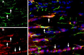
Multiplex staining of IBA1/AIF1 and P2X7 in rat brain. - Immunohistochemical staining of rat corpus callosum (CC) free floating frozen sections using Anti-IBA1/AIF1 Antibody (#ACS-010), (1:1000) and Anti-P2X7 Receptor-ATTO Fluor-550 Antibody (#APR-004-AO) (1:60). A. IBA1/AIF1 immunoreactivity (green) appears in microglia (arrows). B. P2X7 immunostaining (red) appears in microglia (up-pointing arrows) and in other cell types in the corpus callosum (down-pointing arrows). C. Merged image of panels A and B demonstrates partial colocalization of both proteins. Nuclei are demonstrated using DAPI as the counterstain (blue).
|
|
|
|
Multiplex staining of IBA1/AIF1 and P2X7 in rat brain. - Immunohistochemical staining of rat corpus callosum (CC) free floating frozen sections using Anti-IBA1/AIF1 Antibody (#ACS-010), (1:1000) and Anti-P2X7 Receptor-ATTO Fluor-550 Antibody (#APR-004-AO) (1:60). A. IBA1/AIF1 immunoreactivity (green) appears in microglia (arrows). B. P2X7 immunostaining (red) appears in microglia (up-pointing arrows) and in other cell types in the corpus callosum (down-pointing arrows). C. Merged image of panels A and B demonstrates partial colocalization of both proteins. Nuclei are demonstrated using DAPI as the counterstain (blue).
|
|
|
| メーカー |
品番 |
包装 |
|
ALO
|
APR-004-AO
|
50 UL
|
※表示価格について
| 当社在庫 |
なし
|
| 納期目安 |
約10日
|
| 法規制 |
毒・安
|
| 保存温度 |
-20℃
|
|

