|
※サムネイル画像をクリックすると拡大画像が表示されます。
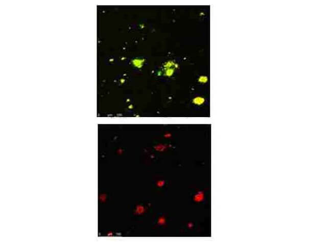
Immunofluorescence Microscopy of Rabbit Anti-beta Amyloid pyro E3 antibody. Tissue: human brain section. Fixation: 0.5% PFA. Antigen retrieval: not required. Primary antibody: beta Amyloid pyro E3 antibody at 5 μg/mL for 1 h at RT. Secondary antibody: Rabbit secondary antibody at 1:10,000 for 45 min at RT. Localization: beta Amyloid pyro E3 is nuclear and cytoplasmic. Staining: Top: s Amyloid pyro E3 as green fluorescent signal, s Amyloid 3 as yellow signal; and Bottom: s Amyloid 3 as red signal with co-incubation of s Amyloid pyro E3 peptide.
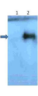
Western Blot of Rabbit Anti-beta Amyloid pyro E3 antibody. Lane 1: beta Amyloid 3 peptide Lane 2: beta Amyloid pyro E3 peptide. Load: 1 μg per lane. Primary antibody: s Amyloid pyro E3 antibody at 2μg/mL for overnight at 4°C. Secondary antibody: IRDye800? rabbit secondary antibody at 1:10,000 for 45 min at RT. Block: 5% BLOTTO overnight at 4°C. Predicted/Observed size: 86.9 kDa for beta Amyloid pyro E3 peptide.
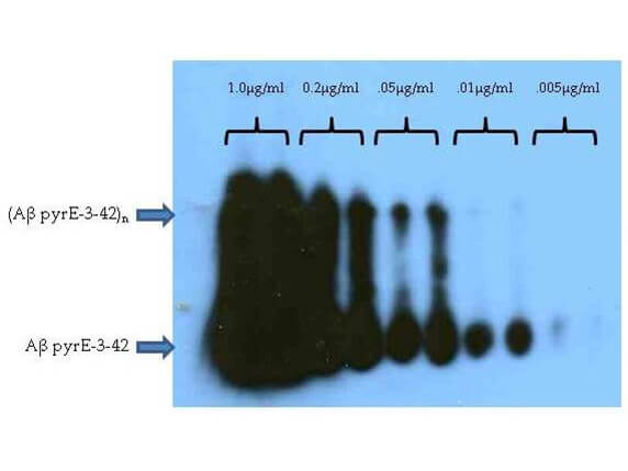
Western Blot of Rabbit Anti-beta Amyloid pyro E3 antibody. Lane 1-10: beta Amyloid pyro E3 peptide. Load: 1 μg per lane 1-2, 0.2 μg per lane 3-4, 0.05 μg per lane 5-6, 0.01 μg per lane 7-8, 0.005 μg per lane 9-10. Primary antibody: s Amyloid pyro E3 antibody at 0.5 μg/mL for overnight at 4°C. Secondary antibody: IRDye800? rabbit secondary antibody at 1:10,000 for 45 min at RT. Block: 5% BLOTTO overnight at 4°C. Predicted/Observed size: 86.9 kDa for beta Amyloid pyro E3 peptide. Cross reactivity with s Amyloid 3 peptide is >1000 fold below pyrE3 reactivity and contains no reactivity towards the s Amyloid 42 Aβ peptide (data not shown).
|
|
|
|
Immunofluorescence Microscopy of Rabbit Anti-beta Amyloid pyro E3 antibody. Tissue: human brain section. Fixation: 0.5% PFA. Antigen retrieval: not required. Primary antibody: beta Amyloid pyro E3 antibody at 5 μg/mL for 1 h at RT. Secondary antibody: Rabbit secondary antibody at 1:10,000 for 45 min at RT. Localization: beta Amyloid pyro E3 is nuclear and cytoplasmic. Staining: Top: s Amyloid pyro E3 as green fluorescent signal, s Amyloid 3 as yellow signal; and Bottom: s Amyloid 3 as red signal with co-incubation of s Amyloid pyro E3 peptide.
|
|
| 別品名 |
rabbit anti-Beta Amyloid pyro E3 Antibody, β-amyloid pyro E3, Alzheimer disease amyloid protein, Beta amyloid, A-beta, ABPP, APPI
|
| 交差種 |
Human
|
| 適用 |
Western Blot
Enzyme Linked Immunosorbent Assay
Immuno Fluorescence
Dot Blot
|
| 免疫動物 |
Rabbit
|
| 抗原部位 |
N-terminus
|
| 標識物 |
Unlabeled
|
| 精製度 |
Affinity Purified
|
| GENE ID |
351
|
| Accession No.(Gene/Protein) |
NP_000475.1, P05067
|
| Gene Symbol |
APP
|
| 参考文献 |
Schilling S, Lauber T, Schaupp M, Manhart S, Scheel E, Bohm G, Demuth HU. On the seeding and oligomerization of pGlu-amyloid peptides (in vitro). Biochemistry;45(41):12393-9. 2006 Oct 17. [PMID: 17029395] He W, Barrow CJ. The A beta 3-pyroglutamyl and 11-pyroglutamyl peptides found in senile plaque have greater beta-sheet forming and aggregation propensities in vitro than full-length A beta. Biochemistry;38(33):10871-7 . 1999 Aug 17. [PMID: 10451383]
|
| [注意事項] |
濃度はロットによって異なる可能性があります。メーカーDS及びCoAからご確認ください。
|
|
| メーカー |
品番 |
包装 |
|
RKL
|
600-401-J14
|
50 UG
|
※表示価格について
| 当社在庫 |
なし
|
| 納期目安 |
約10日
|
| 保存温度 |
-20℃
|
|
※当社では商品情報の適切な管理に努めておりますが、表示される法規制情報は最新でない可能性があります。
また法規制情報の表示が無いものは、必ずしも法規制に非該当であることを示すものではありません。
商品のお届け前に最新の製品法規制情報をお求めの際はこちらへお問い合わせください。
|
※当社取り扱いの試薬・機器製品および受託サービス・創薬支援サービス(納品物、解析データ等)は、研究用としてのみ販売しております。
人や動物の医療用・臨床診断用・食品用としては、使用しないように、十分ご注意ください。
法規制欄に体外診断用医薬品と記載のものは除きます。
|
|
※リンク先での文献等のダウンロードに際しましては、掲載元の規約遵守をお願いします。
|
|
※CAS Registry Numbers have not been verified by CAS and may be inaccurate.
|
