
| 別品名 |
AKT 1 antibody, AKT-1, PKB antibody, PKB gamma antibody, PKBGAMMA antibody, PRKBG antibody, Protein kinase Akt 1 antibody, Protein kinase B gamma antibody, RAC-gamma serine/threonine-protein kinase, RAC-PK-gamma
|
| 種由来 |
Human
|
| 標識物 |
Unlabeled
|
| 精製度 |
Ig fraction - Protein A
|
| 適用 |
Western Blot
Enzyme Linked Immunosorbent Assay
Flow Cytometry
|
| 免疫動物 |
Mouse
|
| 抗体クラス |
IgG2aκ
|
| クローン |
14E5.A2.B2.H9
|
| 交差種 |
Human
Mouse
|
| Accession No.(Gene/Protein) |
P31749
|
| Gene Symbol |
AKT1
|
| 形状 |
滅菌済み液状品
|
| 参考文献 |
[Pub Med ID]30377371
|
| [注意事項] |
濃度はロットによって異なる可能性があります。メーカーDS及びCoAからご確認ください。
|
|
※サムネイル画像をクリックすると拡大画像が表示されます。
Flow Cytometry of Mouse anti AKT1 antibody. Cells: LNCap Cells. Stimulation: none. Primary antibody: Allophycocyanin AKT1 antibody at 1.0 ug/mL for 20 min at 4C.
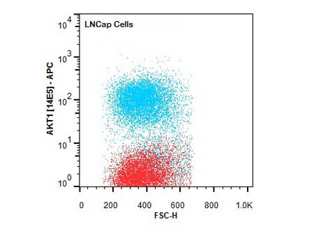
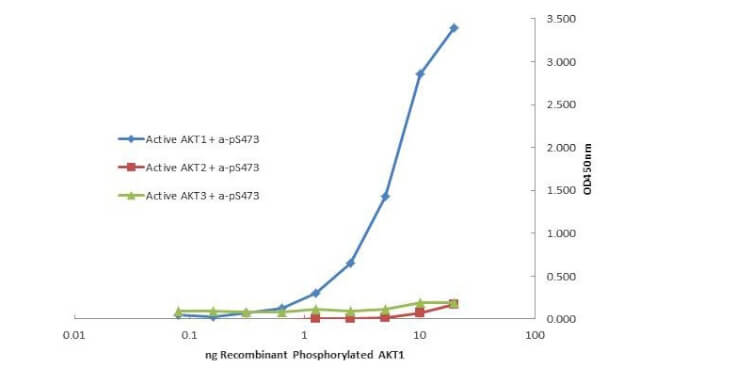
Plate was coated with monoclonal anti AKT1 antibody (capture antibody) followed by incubation with recombinant AKT1 (p/n 009-001-P21), AKT2 (p/n 009-001-P22), AKT3 (p/n 009-001-P23) proteins. Binding was detected with biotinylated monoclonal anti-AKT pS473. The signal shows specificity of the monoclonal anti-AKT1 antibody to recombinant isoform AKT1 protein and not the isoform 2 and 3.
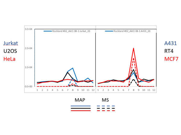
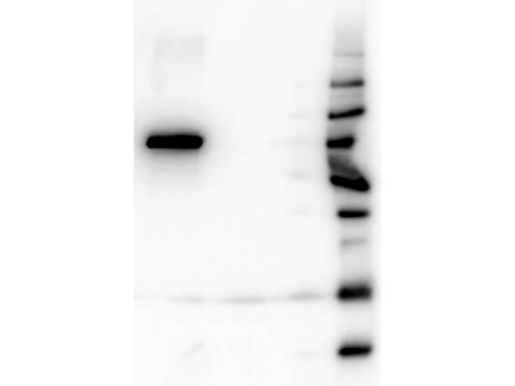
PAGE-MAP (microsphere affinity proteomics) of Mouse Anti-AKT1 Antibody. (Catalog Number: 200-301-I51, Lot Number: 29014). Antibody array western blot binding of gelfree size separated fractions of multiple lysates (solid lines) and shotgun mass spectroscopy identification (dashed lines) of the target band run in parallel correlate confirming the specificity of this antibody against AKT1. Data was provided by the Lund-Johansen lab of Oslo University Hospital. For more information on PAGE-MAP/IP-MS identification of antibody specificity and its large-scale implementation for antibody validation see Sikorski et. al., (2018) Nature Methods 15, 909-912.
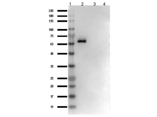
Western Blot of Mouse Anti-AKT1 Antibody. Lane 1: Opal Prestained Molecular Weight Protein (p/n MB-210-0500). Lane 2: AKT1 protein (p/n 009-001-P21). Lane 3: AKT2 protein (p/n 009-001-P22). Lane 4: AKT3 protein (p/n 009-001-P23). Load: 50ng. Blocking: BlockOut Buffer (p/n MB-073) for 30 min at RT. Primary Antibody: Anti-AKT1 at 1ug/mL o/n at 4C. Secondary Antibody: Rabbit Anti-Mouse IgG HRP (p/n 610-403-C46, Lot 20121) at 1:40,000 in MB-073 for 30 min at RT.
Western Blot of Mouse Anti-AKT1 antibody. Lane 1: GST Tagged recombinant AKT1. Lane 2: GST Tagged recombinant AKT2. Lane 3: GST Tagged recombinant AKT3. Load: 25 ng per lane. Primary antibody: AKT1 antibody at 1:1,000 for overnight at 4C. Secondary antibody: Peroxidase mouse secondary antibody at 1:40,000 for 30 min at RT. Block: MB-070 for 30 min at RT. Predicted/Observed size: 78 kDa for AKT1. Other band(s): none.
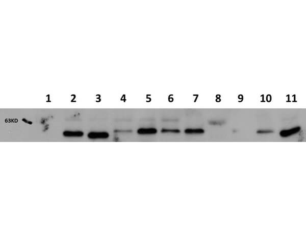
Western Blot of Mouse Anti-AKT1 antibody. Lane 1: AKT-1 Null. Lane 2: WT. Lane 3: MEF #1. Lane 4 : A549 (p/n W09-001-GX4). Lane 5: Calu-1. Lane 6: PC-3 (p/n W09-001-GV6). Lane 7: HepG2 (p/n W09-001-GJ5). Lane 8: Jurkat (p/n W09-001-370). Lane 9: SKOV3 (p/n W09-001-GX9). Lane 10: HEK293T (p/n W09-001-GX5). Lane 11: C2C12 (p/n W10-001-GL7). Load: 20 ug per lane. Primary antibody: AKT1 antibody at 1:1,000 for overnight at 4C. Secondary antibody: Peroxidase mouse secondary antibody at 1:40,000 for 30 min at RT. Block: MB-070 for 30 min at RT. Predicted/Observed size: 56 kDa for AKT1. Other band(s): none.
|
|
|
|
Flow Cytometry of Mouse anti AKT1 antibody. Cells: LNCap Cells. Stimulation: none. Primary antibody: Allophycocyanin AKT1 antibody at 1.0 ug/mL for 20 min at 4C.
|
|
|
| メーカー |
品番 |
包装 |
|
RKL
|
200-301-I51L
|
1 MG
|
※表示価格について
| 当社在庫 |
なし
|
| 納期目安 |
約10日
|
| 保存温度 |
-20℃
|
|

