|
※サムネイル画像をクリックすると拡大画像が表示されます。
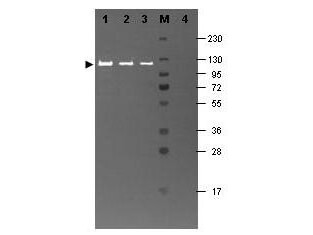
Western blotting using Rockland's Fluorescein conjugated anti-b-Galactosidase antibody shows a band at ~117 kDa (lanes 1 - 3) corresponding to 60 ng, 30 ng and 15 ng, respectively of b-Gal present in partially purified preparations (arrowhead). Lane 4 shows no cross reactivity with proteins present in a non-specific control E.coli lysate. Proteins were resolved on a 4-20% Tris-Glycine gel by SDS-PAGE and transferred to nitrocellulose and blocking using Blocking Buffer for Fluorescent Western Blotting (p/n MB-070). The membrane was probed with fluorescein conjugated anti-b-Galactosidase (p/n 200-4236) diluted to 1:10,000. Reaction occurred for 2 hours at room temperature. Molecular weight estimation was made by comparison to a prestained MW marker in lane M. Fluorescence image was captured using the VersaDoc(R) Imaging System developed by BIO-RAD. Other detection systems will yield similar results
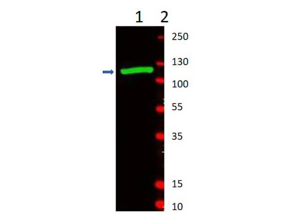
Western Blot of Rabbit Anti-Beta-Galactosidase Antibody. Lane 1: partially purified preparation b-Galactosidase [1.0μg]. Lane 2: Molecular Weight Marker. Primary Antibody: Anti-Beta-Galactosidase at 1:1000 overnight at 2-8°C. Secondary Antibody: Goat Anti-Rabbit IgG IRDye(R)800 (p/n 611-132-122) 1:10,000 for 45min at RT. Observed MW: ~117kDa.
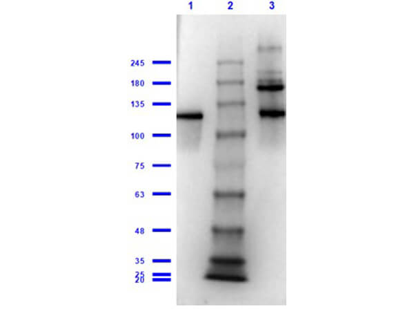
Western Blot of Rabbit Anti-Beta-Galactosidase Antibody. Lane 1: Beta-Galactosidase Reduced [0.1μg]. Lane 2: Opal Prestained Molecular Weight Marker (p/n MB-210-0500). Lane 3: Beta-Galactosidase Non-Reduced [0.1μg]. Primary Antibody: Anti-Beta-Galactosidase at 1:1000 overnight at 2-8°C. Secondary Antibody: Goat Anti-Rabbit IgG HRP (p/n 611-103-122) 1:70,000 for 30min at RT. Expected MW: ~117kDa.
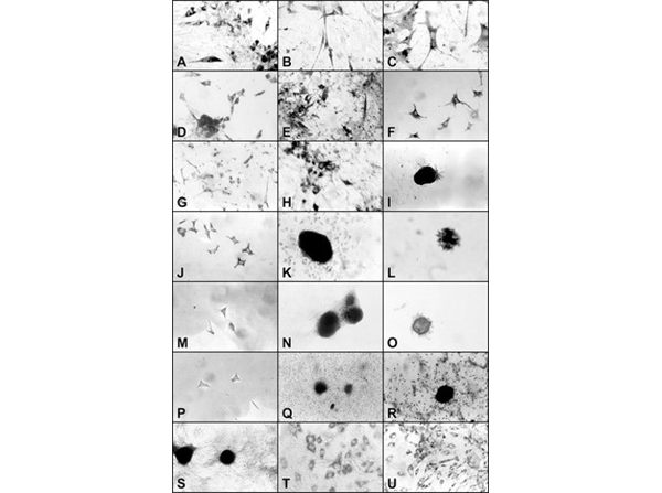
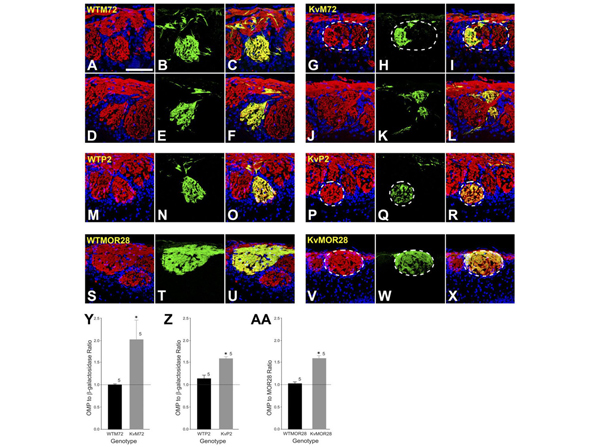
Absence of Kv1.3 ion channel induces the development of heterogeneous glomeruli.?A?L:?Representative two-photon confocal micrographs of WTM72 (left panels; A?F) and KvM72 glomeruli (right panels; G?L). A,D,G,J: Red channel; sections were processed for olfactory marker protein (OMP; 1:1,000) immunocytochemistry to visualize axons projecting from all mature OSNs. B,E,H,K: Green channel; sections were processed for β-galactosidase immunocytochemistry (1:1,000). C,F,I,L: Merged image, yellow. Note the incomplete innervation of the M72 glomerulus (I,L) in KvM72 as evidenced by red only fibers creating a heterogeneous M72 glomerulus. Postnatal day 20.?M?R:?Representative two-photon confocal micrograph of a WTP2 (M?O) and a KvP2 glomerulus (P?R). Red channel represents OMP; green, β-galactosidase; yellow, merged image. Note the presence of a heterogeneous P2 glomerulus (R) in the KvP2 mouse. A few OMP-positive, β-galactosidase-negative fibers are also visible in WTP2 mice (O).?S?X:?Representative two-photon confocal micrograph of a WTMOR28 (S-U) and a Kv-MOR28 glomerulus (V?X). Red channel represents OMP; green, MOR28 immunoreactivity; yellow, merged image. KvMOR28 mice exhibit heterogeneous glomeruli (X).?Y:?Histogram plot of αOMP to α-β-galactosidase pixel density for M72 glomeruli of WTM72 and KvM72 mice. A ratio of 1.0 represents complete overlapping of OMP and β-galactosidase-immunoreactive axons (homogeneous glomerulus), whereas a ratio > 1.0 represents more OMP-immunoreactive axons (heterogeneous glomeruli). Values are mean ± SEM. *, significantly different by Student’s?t-test,?p?? 0.05.?Z:?Same as Y but for WTP2 and KvP2.?AA:?Histogram plot of αOMP to αMOR28 pixel density for MOR28 glomeruli of WTMOR28 vs. KvMOR28 mice. Values are mean ± SEM. *, significantly different by Student’s?t-test,?p?? 0.05. Scale bar = 100 μm in A (applies to A?X). Fig 5. PMID:?18022950
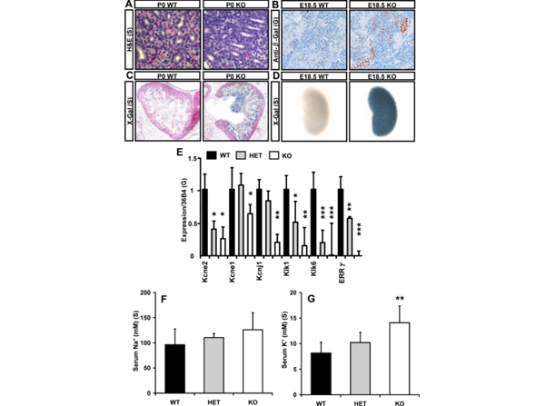
Loss of ERRγ alters renal markers. A, Hematoxylin and eosin staining of kidney does not reveal significant differences in ERRγ null mice. B, Anti-β-galactosidase staining indicates ERRγ expression in the distal tubules. C, X-gal staining of frozen sections shows that ERRγ is expressed in the distal tubules and collecting ducts of the kidney. D, Whole-mount X-gal staining reveals the high expression of ERRγ throughout the cortex of the kidney. E, In kidney, loss of ERRγ produces a down-regulation of potassium channels and components of the KSS as detected by triplicate quantitative PCR (n = 3 WT; 9 HET; 6 KO). F, Serum sodium levels did not increase to statistically significant degree in ERRγ null mice at E18.5. G, Serum potassium levels were increased by more than 70% in ERRγ null animals at E18.5. Values ±?sd; *,?P?< 0.05; **,?P?< 0.01; ***,?P?< 0.001 by two-tailed Student's?t?test. S, Salk mice; G, GlaxoSmithKline mice; H&E, hematoxylin and eosin; HET, heterozygous. Fig 5. PMID:?19965931
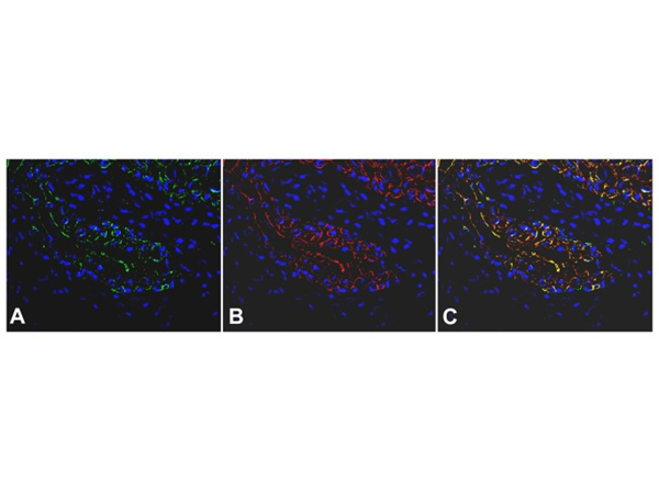
Immunofluorescent staining of β-galactosideasae and cytokeratin expression of urinary bladder after intravesical treatment with Adeno-Cre. Adeno-Cre (8.46×10⁸?pfu per mouse) was delivered into the bladder lumen of ROSA mice for 2 hours (n=5). Mice were sacrificed and their bladders were removed and processed for immunofluorescence staining. The urothelial mucosa shows in green β-galactosidase (A) and in red cytokeratin 7 expressions (B). In Figure (C), overlapping of the fields (A) and (B) demonstrates the co-localization of β-galactosidase and cytokeratin 7 expressions (orange-yellow). Nuclei were stained with DAPI (blue color). β-galactosidase expression is scattered and observed mainly in the 2/3 upper (inner) layer of the urothelium versus cytokeratin expression which is evenly present in the full thickness of the urothelium. Fig 3. PMID:?24058630
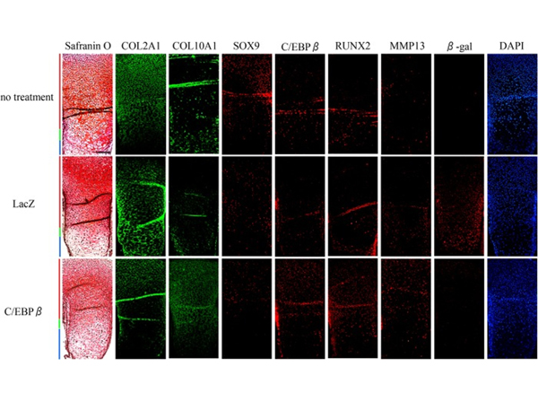
C/EBPβ repressed the expression of COL2A1 and SOX9 in?ex vivo?organ cultures.?Ex vivo?organ culture of tibias dissected from E14.5 mouse embryos are shown. Tibias were cultured for 4 days without adenovirus vectors (top row) or after transfection with adenovirus vectors expressing LacZ control (middle row) or C/EBPβ (bottom row). Safranin O staining and immunofluorescent staining were performed to localize COL2A1, COL10A1, SOX9, C/EBPβ, RUNX2, and MMP13. β-Galactosidase antibody (β-gal) was used to confirm the transfection efficiency of adenovirus vectors. DAPI was used as a counterstain.?Red,?green, and?blue bars?indicate the proliferative, prehypertrophic, and hypertrophic zones, respectively.?Scale bar, 500 μm. Histological analysis was repeated at least twice for each sample from six pairs of limbs, respectively. Fig 7. PMID:?24344131
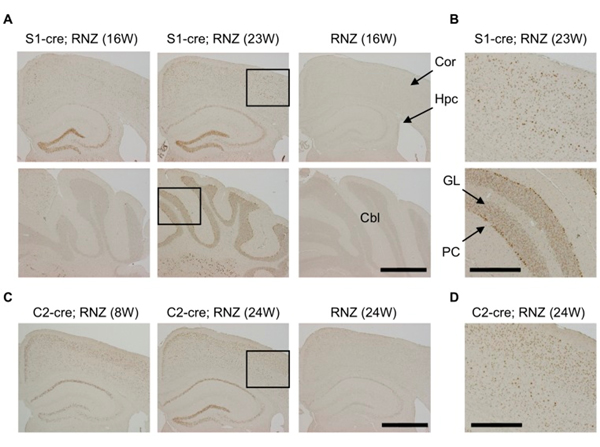
Transgenic mice for synapsinI-cre (S1-cre) or camk2a-cre (C2-cre) were crossed with RNZ mice. RNZ male mice harboring S1-cre or C2-cre at indicated weeks of age were subjected to anti-LacZ staining to detect cre-mediated DNA recombination. RNZ mice without a cre transgene were used as controls. (A) In S1-cre; RNZ mice at 16 weeks of age, LacZ-positive cells were strongly detected in dentate gyrus and CA3 in hippocampus and some cortical cells, but very few were seen in cerebellum. At 23 weeks, LacZ expression became wider in the cortex and hippocampus, and was clearly detected in cerebellum. No distinct LacZ expression was detected in the control RNZ mice. (B) High magnification of boxed region in (A). LacZ expression was broadly detected in multiple layers of cortex and Purkinje and granular cells of cerebellum in 23 week-old S1-cre; RNZ mice. (C) LacZ-positive cells were broadly detected in brains of 8-week-old C2-cre; RNZ mice, especially in layer II/III of cortex and CA1 of hippocampus. It became wider at 24 weeks of age. Again, no distinct LacZ expression was observed in the control RNZ mice. (D) High magnification of boxed region in (C), indicating detection of LacZ-positive cells in multiple layers of cortex in 24 week-old C2-cre; RNZ mice. Cor (cortex), Hpc (hippocampus), Cbl (cerebellum), PC (Purkinje cell) and GL (granular layer). Bars are 1 mm (A, C) and 0.4 mm (B, D). Fig 1. PMID:?24391875
|
|
|
|
Western blotting using Rockland's Fluorescein conjugated anti-b-Galactosidase antibody shows a band at ~117 kDa (lanes 1 - 3) corresponding to 60 ng, 30 ng and 15 ng, respectively of b-Gal present in partially purified preparations (arrowhead). Lane 4 shows no cross reactivity with proteins present in a non-specific control E.coli lysate. Proteins were resolved on a 4-20% Tris-Glycine gel by SDS-PAGE and transferred to nitrocellulose and blocking using Blocking Buffer for Fluorescent Western Blotting (p/n MB-070). The membrane was probed with fluorescein conjugated anti-b-Galactosidase (p/n 200-4236) diluted to 1:10,000. Reaction occurred for 2 hours at room temperature. Molecular weight estimation was made by comparison to a prestained MW marker in lane M. Fluorescence image was captured using the VersaDoc(R) Imaging System developed by BIO-RAD. Other detection systems will yield similar results
|
|
| 別品名 |
rabbit anti-Beta Galactosidase Antibody, rabbit anti-beta gal antibody, β-Gal, Anti-β-Gal Antibody
|
| 別包装 |
あり
|
| 適用 |
Western Blot
Enzyme Linked Immunosorbent Assay
|
| 免疫動物 |
Rabbit
|
| 標識物 |
Unlabeled
|
| 精製度 |
Ig fraction - Ion Exchange /Gel Filtration
|
| Accession No.(Gene/Protein) |
NP_414878.1, P00722
|
| Gene Symbol |
lacZ
|
| Tag情報 |
b-GAL
|
| 参考文献 |
[[Cosmo Reference Collection]][Pub Med ID]14983513,15890684,16793894,18022950,19965931,21653538,24058630,24344131,24391875,24566496,25218638,27687130,28754980,32020011,15602772,31916371,27867035
|
| [注意事項] |
濃度はロットによって異なる可能性があります。メーカーDS及びCoAからご確認ください。
|
|
| メーカー |
品番 |
包装 |
|
RKL
|
200-4136-0100
|
100 UG
|
※表示価格について
| 当社在庫 |
なし
|
| 納期目安 |
約10日
|
| 法規制 |
毒
|
| 保存温度 |
4℃
|
|


