|
※サムネイル画像をクリックすると拡大画像が表示されます。
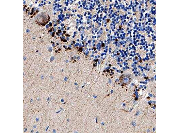
Immunohistochemistry analysis of Anti-DR5/TRAIL R2/TNFRSF10B antibody at 1:200 concentration, formalin fixed paraffin-embedded (FFPE) human brain cerebellum, on a Bond Rx autostainer (Leica Biosystems). The assay involved 30 minutes of heat induced antigen retrieval (HIER) using 10mM sodium citrate buffer (pH 9.0) and endogenous peroxidase quenching with peroxide block. The sections were incubated with primary antibody for 15 minutes and Bond Polymer Refine Detection (Leica Biosystems) with DAB was used for signal development followed by counterstaining with hematoxylin. Cytoplasmic staining was observed in the Purkinje cell layer.
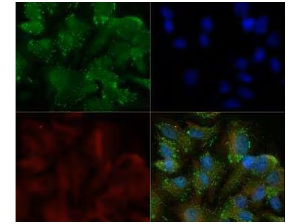
Immunocytochemistry/Immunofluorescence analysis of Anti-DR5/TRAIL R2/TNFRSF10B Antibody. HepG2 cells were fixed for 10 minutes using 10% formalin and then permeabilized for 5 minutes using 1X TBS + 0.5% Triton-X100. The cells were incubated with anti-DR5 at 5ug/ml overnight at 4°C and detected with an anti-rabbit Dylight 488 (Green) at a 1:500 dilution. Alpha tubulin (DM1A) was used as a co-stain at a 1:1000 dilution and detected with an anti-mouse Dylight 550 (Red) at a 1:500 dilution. Nuclei were counterstained with DAPI (Blue). Cells were imaged using a 40X objective.
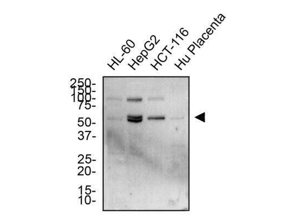
Western Blot analysis of Anti-DR5/TRAILR2/TNFRSF10B Antibody. Total protein from HL-60, HepG2, HCT-116 and human placenta was separated on a 12% gel by SDS-PAGE, transferred to PVDF membrane and blocked in 5% non-fat milk in TBST. The membrane was probed with 2.0 ug/ml anti-DR5 in 1% non-fat milk in TBST and detected with an anti-rabbit HRP secondary antibody using chemiluminescence.
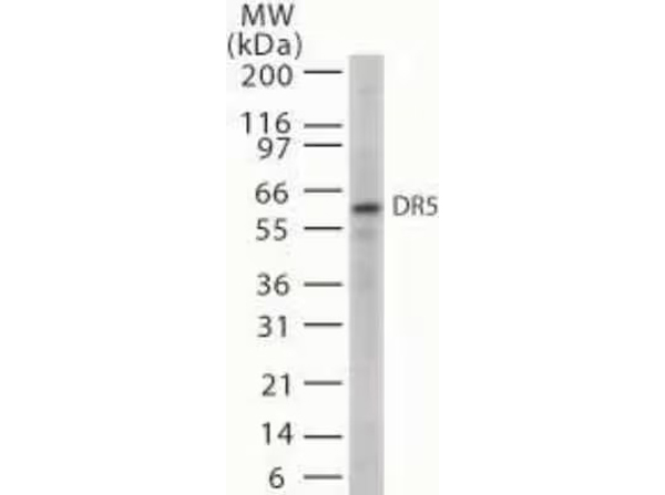
Western Blot analysis of Anti-DR5/TRAILR2/TNFRSF10B Antibody. Detection of 20 ug of whole cell lysates from HL60 cells with anti-D5 at 5μg/ml.
|
|
|
|
Immunohistochemistry analysis of Anti-DR5/TRAIL R2/TNFRSF10B antibody at 1:200 concentration, formalin fixed paraffin-embedded (FFPE) human brain cerebellum, on a Bond Rx autostainer (Leica Biosystems). The assay involved 30 minutes of heat induced antigen retrieval (HIER) using 10mM sodium citrate buffer (pH 9.0) and endogenous peroxidase quenching with peroxide block. The sections were incubated with primary antibody for 15 minutes and Bond Polymer Refine Detection (Leica Biosystems) with DAB was used for signal development followed by counterstaining with hematoxylin. Cytoplasmic staining was observed in the Purkinje cell layer.
|
|
| 別品名 |
Tumor necrosis factor receptor superfamily member 10B, TNFRSF10B, DR5, KILLER, TRAILR2, TRICK2, ZTNFR9, Death receptor 5, TNF-related apoptosis-inducing ligand receptor 2, TRAIL receptor 2, TRAIL-R2, CD_antigen=CD262
|
| 交差種 |
Human
|
| 適用 |
Western Blot
Immunohistochemistry
Immuno Fluorescence
Flow Cytometry
|
| 免疫動物 |
Rabbit
|
| 抗体クラス |
IgG
|
| 抗原部位 |
C-terminus
|
| 標識物 |
Unlabeled
|
| 精製度 |
Ig fraction - Protein G
|
| GENE ID |
8795
|
| Accession No.(Gene/Protein) |
NP_003833.4, O14763
|
| Gene Symbol |
TNFRSF10B
|
| 参考文献 |
1. Pan G, Ni J, Wei YF, Yu G, Gentz R, Dixit VM. An antagonist decoy receptor and a death domain-containing receptor for TRAIL. Science 1997;277:815-8 2. Sheridan JP, Marsters SA, Pitti RM, Gurney A, Skubatch M, Baldwin D, Ramakrishnan L, Gray CL, Baker K, Wood WI, Goddard AD, Godowski P, Ashkenazi A. Control of TRAIL-induced apoptosis by a family of signaling and decoy receptors. Science 1997;277:818-21
|
| [注意事項] |
濃度はロットによって異なる可能性があります。メーカーDS及びCoAからご確認ください。
|
|
| メーカー |
品番 |
包装 |
|
RKL
|
200-401-H72
|
100 UG
|
※表示価格について
| 当社在庫 |
なし
|
| 納期目安 |
約10日
|
| 保存温度 |
-20℃
|
|
※当社では商品情報の適切な管理に努めておりますが、表示される法規制情報は最新でない可能性があります。
また法規制情報の表示が無いものは、必ずしも法規制に非該当であることを示すものではありません。
商品のお届け前に最新の製品法規制情報をお求めの際はこちらへお問い合わせください。
|
※当社取り扱いの試薬・機器製品および受託サービス・創薬支援サービス(納品物、解析データ等)は、研究用としてのみ販売しております。
人や動物の医療用・臨床診断用・食品用としては、使用しないように、十分ご注意ください。
法規制欄に体外診断用医薬品と記載のものは除きます。
|
|
※リンク先での文献等のダウンロードに際しましては、掲載元の規約遵守をお願いします。
|
|
※CAS Registry Numbers have not been verified by CAS and may be inaccurate.
|
