
| 別品名 |
Toll-like receptor 3, CD283
|
| 種由来 |
Human
|
| 標識物 |
Unlabeled
|
| 精製度 |
Ig fraction - Protein G
|
| 適用 |
Western Blot
Immunohistochemistry
Immuno Fluorescence
Flow Cytometry
Immunoprecipitation
|
| 免疫動物 |
Mouse
|
| 抗体クラス |
IgG1κ
|
| クローン |
40C1285.6
|
| 交差種 |
Human
|
| GENE ID |
7098
|
| Accession No.(Gene/Protein) |
NP_003256, O15455
|
| Gene Symbol |
TLR3
|
| 形状 |
液状
|
| [注意事項] |
濃度はロットによって異なる可能性があります。メーカーDS及びCoAからご確認ください。
|
|
※サムネイル画像をクリックすると拡大画像が表示されます。
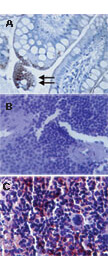
Immunohistochemistry of mouse Anti TLR3 antibody. Tissue A: Human gut lumen (longitudinal section, tranverse region) using TLR3. Tissue B: Mouse spleen tissue using isotype control. Tissue C: Mouse spleen tissue using TLR3. Fixation: formalin fixed paraffin embedded. Antigen retrieval: not required. Primary antibody: TLR3 antibody at tissue A at 10 ug/mL and at tissue C at 5 mg/ml for 1 h at RT. Secondary antibody: Peroxidase mouse secondary antibody at 1:10,000 for 45 min at RT. Localization: TLR3 is an endoplasmic reticulum membrane and a single pass type 1 membrane protein. Staining: TLR3 is precipitated as a red signal with hematoxylin purple nuclear counterstain.
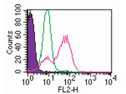
Flow Cytometry of Mouse Anti-TLR3 antibody. Cells: Human monocytes. Stimulation: none. Primary Antibody: Anti-TLR3 antibody at 0.5 ug (red) and isotype control (green). Secondary Antibody: Goat anti-mouse IgG1 PE conjugate (BD).
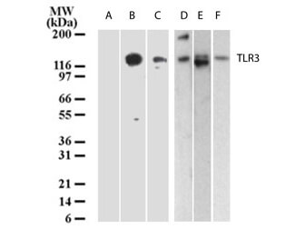
Western Blot of Mouse Anti-TLR3 antibody. Lane A: untransfected 293. Lane B: 293 cells with human TLR3 cDNA. Lane C: human intestine. Lane D: placenta. Lane E: heart. Lane F: ovary. Primary antibody: TLR3 antibody at 3 ug/mL for overnight at 4C. Secondary antibody: Goat anti-mouse HRP conjugate at 1:10,000 for 45 min at RT. Block: 5% BLOTTO overnight at 4C. Predicted/Observed size: 80 kDa for TLR3. Other band(s): none.
|
|
|
|
Immunohistochemistry of mouse Anti TLR3 antibody. Tissue A: Human gut lumen (longitudinal section, tranverse region) using TLR3. Tissue B: Mouse spleen tissue using isotype control. Tissue C: Mouse spleen tissue using TLR3. Fixation: formalin fixed paraffin embedded. Antigen retrieval: not required. Primary antibody: TLR3 antibody at tissue A at 10 ug/mL and at tissue C at 5 mg/ml for 1 h at RT. Secondary antibody: Peroxidase mouse secondary antibody at 1:10,000 for 45 min at RT. Localization: TLR3 is an endoplasmic reticulum membrane and a single pass type 1 membrane protein. Staining: TLR3 is precipitated as a red signal with hematoxylin purple nuclear counterstain.
|
|
|
| メーカー |
品番 |
包装 |
|
RKL
|
200-301-I24
|
100 UG
|
※表示価格について
| 当社在庫 |
なし
|
| 納期目安 |
約10日
|
| 保存温度 |
-20℃
|
|

