
| 別品名 |
IKBA, MAD3, NFKBI, NF-kappa-B inhibitor alpha, I-kappa-B-alpha, IkB-alpha, IkappaBalpha, Major histocompatibility complex enhancer-binding protein MAD3
|
| 種由来 |
Human
|
| 標識物 |
Unlabeled
|
| 精製度 |
Ig fraction - Protein G
|
| 適用 |
Western Blot
Immunohistochemistry
Immuno Fluorescence
Flow Cytometry
Immunoprecipitation
Chromatin Immunoprecipitation
|
| 免疫動物 |
Mouse
|
| 抗体クラス |
IgG1κ
|
| クローン |
6A920
|
| 交差種 |
Human
Mouse
|
| GENE ID |
4792
|
| Accession No.(Gene/Protein) |
NP_065390, P25963
|
| Gene Symbol |
FKBIA
|
| 形状 |
液状
|
| 参考文献 |
1. Verma, I.M., Genes. Dev. 9: 2723-2735, 1995. 2. Verma, I. And Stevenson, J.K. Proc. Nat. Acad. Sci USA 94: 11758 (1997). 3. DiDonato, J.A., et al. Nature 388: 548- (1997). 4. Regnier, C.H., et al. Cell 90, 373 (1997). 5. Mercurio, F., et al. Science 278: 860 (1997). 6. Yamaoka, S., et al. Cell 93: 1231-1240 (1998). 7. Sanjo H, Kawai T, and Akira S. DRAKs, novel serine/threonine kinases related to death associated protein kinase that trigger apoptosis. J Biol Chem 273 (44): 29066-29071, 1998. 8. Matsumoto M, Takeda K, Sanjo H, and Akira S. ZIP kinase, a novel serine/threonine kinase which mediates apoptosis. Mol Cell Biol 18 (3): 1642-1651, 1998.
|
| [注意事項] |
濃度はロットによって異なる可能性があります。メーカーDS及びCoAからご確認ください。
|
|
※サムネイル画像をクリックすると拡大画像が表示されます。
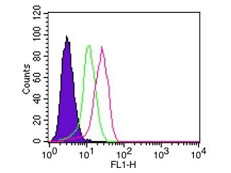
Flow Cytometry of Mouse Anti IkB alpha antibody. Cells: 10^6 ThP 1 cells. Stimulation: none. Primary Antibody: IkBa antibody at 0.25 ug/mL (red) and isotype control (green). Secondary Antibody: anti mouse IgG FITC.
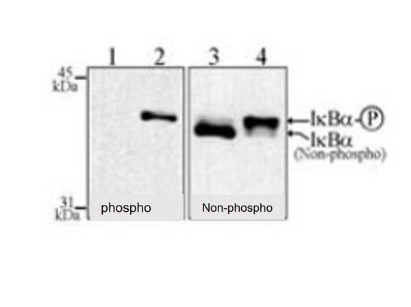
Western Blot of Mouse Anti-IkB alpha antibody. Jurkat cells treated with 00μg/mL pf ALLN for 30 min. Lane 1: Lysate without 1 nM TNF-a. Lane 2: Lysate with 1 nM TNF-a. Lane 3: Lysate without 1 nM TNF-a. Lane 4: Lysate with 1 nM TNF-a. Load: 30 μg per lane. Primary antibody: IkB alpha antibody at 4 μg/mL for overnight at 4°C. Secondary antibody: IRDye800? mouse secondary antibody at 1:10,000 for 45 min at RT. Block: 5% BLOTTO overnight at 4°C. Predicted/Observed size: 35.6 kDa for IkB alpha. Other band(s): both IkB alpha Phoshpo and non-phoshpo.
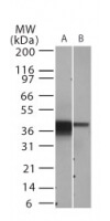
Western Blot of Mouse Anti-IkB alpha antibody. Lane 1: Daudi cells. Lane 2: NIH 3T3 whole cell lysate. Load: 30 ug per lane. Primary antibody: IkB alpha antibody at 2 ug/mL for overnight at 4C. Secondary antibody: IRDye800TM mouse secondary antibody at 1:10,000 for 45 min at RT. Block: 5% BLOTTO overnight at 4C. Predicted/Observed size: 35.6 kDa for IkBa. Other band(s): none.
|
|
|
|
Flow Cytometry of Mouse Anti IkB alpha antibody. Cells: 10^6 ThP 1 cells. Stimulation: none. Primary Antibody: IkBa antibody at 0.25 ug/mL (red) and isotype control (green). Secondary Antibody: anti mouse IgG FITC.
|
|
|
| メーカー |
品番 |
包装 |
|
RKL
|
200-301-H80
|
100 UG
|
※表示価格について
| 当社在庫 |
なし
|
| 納期目安 |
約10日
|
| 保存温度 |
-20℃
|
|

