|
※サムネイル画像をクリックすると拡大画像が表示されます。
Figure 1: Immunohistochemistry on frozen section of human kidney epithelium
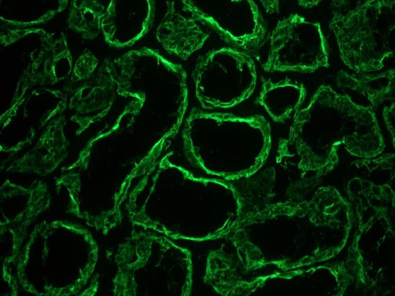
Figure 2: Immunohistochemistry on frozen section of human colon
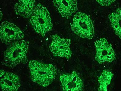
Figure 3: Immunohistochemistry on frozen section of swine liver hepatocytes
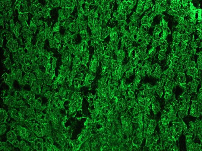
Figure 4: Immunofluorescence staining of epithelial tissues in a 2 days old zebrafish embryo.
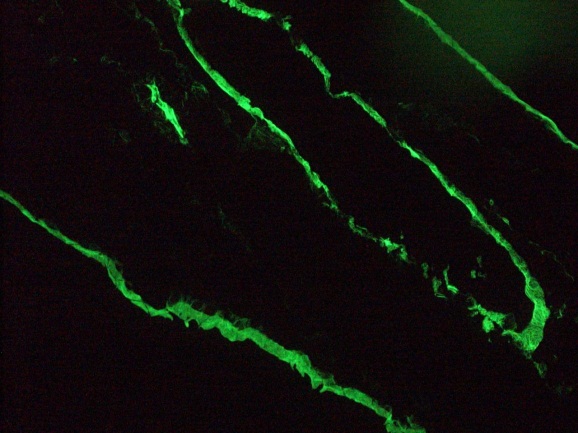
Figure 6: Immunofluorescence staining of a 5 days old zebrafish embryo. Left panel: DAPI-staining of cell nuclei, providing an overview of the tissue section used for immunostaining
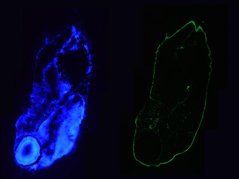
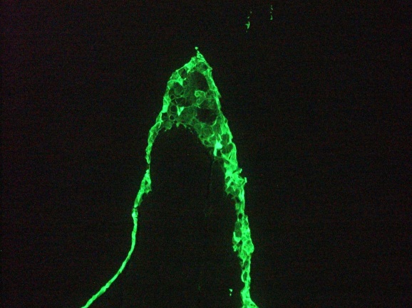
Figure 7: Immunofluorescence staining of 1 month old zebrafish embryo
|
|
|
|
Figure 1: Immunohistochemistry on frozen section of human kidney epithelium
|
|
| 別品名 |
0
|
| 交差種 |
Human
Mouse
Rat
Rabbit
Porcine
Chicken
Canine
Hamster
Zebrafish
|
| 適用 |
Western Blot
IHC frozen section
Immunocytochemistry (cell)
Flow Cytometry
|
| 免疫動物 |
Mouse
|
| クローン |
RGE53
|
| 抗体クラス |
IgG1
|
| 標識物 |
Unlabeled
|
| 精製度 |
Purified
|
| その他 |
[Uniprot ID]P05783
|
| 参考文献 |
1. Ramaekers, F., Huysmans, A., Moesker, O., Kant, A., Jap, P., Herman, C., and Vooijs, P. (1983). Monoclonal antibody to Keratin filaments, specific for glandular epithelia and their tumors. Use in surgical pathology, Lab Invest 49, 353-61.
2. Ramaekers, F. C., Puts, J. J., Moesker, O., Kant, A., Huysmans, A., Haag, D., Jap, P. H., Herman, C. J., and Vooijs, G. P. (1983). Antibodies to intermediate filament proteins in the immunohistochemical identifiCation of Human tumours: an overview, Histochem J 15, 691-713.
3. Puts, J. J., Moesker, O., Kenemans, P., Vooijs, G. P., and Ramaekers, F. C. (1985). Expression of Cytokeratins in early neoplastic epithelial lesions of the uterine cervix, Int J Gynecol Pathol 4, 300-13.
4. Ramaekers, F., van Niekerk, C., Poels, L., Schaafsma, E., Huijsmans, A., Robben, H., Schaart, G., and Vooijs, P. (1990). Use of monoclonal antibodies to Keratin 7 in the differential diagnosis of adenocarcinomas, Am J Pathol 136, 641-55.
5. Raats, J. M., Pieper, F. R., Vree Egberts, W. T., Verrijp, K. N., Ramaekers, F. C., and Bloemendal, H. (1990). Assembly of amino-terminally deleted desmin in vimentin-free cells, J Cell Biol 111, 1971-85.
6. Smedts, F., Ramaekers, F., Robben, H., Pruszczynski, M., van Muijen, G., Lane, B., Leigh, I., and Vooijs, P. (1990). Changing patterns of Keratin expression during progression of cervical intraepithelial neoplasia, Am J Pathol 136, 657-68.
7. Smedts, F., Ramaekers, F., Troyanovsky, S., Pruszczynski, M., Link, M., Lane, B., Leigh, I., Schijf, C., and Vooijs, P. (1992). Keratin expression in cervical cancer, Am J Pathol 141, 497-511.
8. van Leenders, G., Dijkman, H., Hulsbergen-van de Kaa, C., Ruiter, D., and Schalken, J. (2000). Demonstration of intermediate cells during Human prostate epithelial differentiation in situ and in vitro using triple-staining confocal scanning microscopy, Lab Invest 80, 1251-8.
|
|
| メーカー |
品番 |
包装 |
|
NOR
|
MUB0326P
|
100 UG
|
※表示価格について
| 当社在庫 |
なし
|
| 納期目安 |
約10日
|
| 保存温度 |
4℃
|
|
