

| 別品名 |
H4/A, H4FE, H4FG, H4FH, H4FI, H4FJ, H4FK, H4FM, H4FN, H4FO, H4/M, H4/N, H4/O, H4/p, H4F2, H4FA, H4FB, H4FC, H4FD, H4/B, H4/C, H4/D, H4/E, H4/G, H4/H, H4/I, H4/J, H4/K, HIST1H4H, HIST1H4I, HIST1H4J, HIST1H4K, HIST1H4L, HIST2H4, HIST2H4A, HIST2H4B, HIST1H4A, HIST1H4B, HIST1H4C, HIST1H4D, HIST1H4E, HIST1H4F, histone cluster 4, H4, histone 4, H4, histone H4, MGC24116
|
| 抗原部位 |
N-terminus
|
| 種由来 |
Human
|
| 標識物 |
Unlabeled
|
| 精製度 |
Affinity Purified
|
| 適用 |
Western Blot
Immuno Fluorescence
Chromatin Immunoprecipitation
Dot Blot
|
| 免疫動物 |
Rabbit
|
| 抗体クラス |
IgG
|
| 交差種 |
Human
Mouse
Caenorhabditis elegans
|
| 翻訳後修飾 |
メチル化
|
| GENE ID |
121504
|
| Accession No.(Gene/Protein) |
NP_001029249, P62805
|
| Gene Symbol |
HIST4H4
|
| 形状 |
滅菌済み液状品
|
| 参考文献 |
[Pub Med ID]29123096
|
|
※サムネイル画像をクリックすると拡大画像が表示されます。
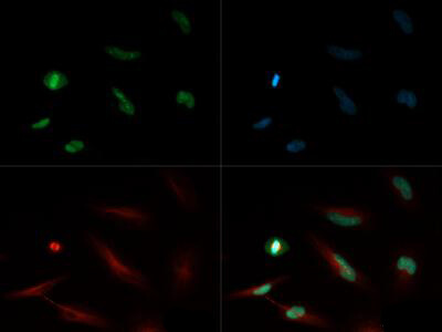
Immunofluorescence of Rabbit Anti Histone H4 [Monomethyl Lys20] Antibody. Tissue: HeLa cells. Fixation: 0.5% PFA. Antigen retrieval: Not required. Primary antibody: Histone H4 [Monomethyl Lys20] antibody at a 1:500 dilution for 1 h at RT. Secondary antibody: FITC secondary antibody at 1:10,000 for 45 min at RT. Localization: Histone H4 [Monomethyl Lys20] is nuclear and chromosomal. Staining: Histone H4 [Monomethyl Lys20] is expressed in green, nuclei and alpha tubulin are counterstained with DAPI (blue) and Dylight 550 (red).
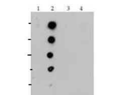
Dot Blot of Rabbit Histone H4 [Monomethyl Lys20] Antibody. Antigen: Lane 1: unmodified. Lane 2: K20me1. Lane 3: K20me2. Lane 4: K20me3. Load: 0.6, 1, 3, 6, and 10 picomoles of peptide. Primary antibody: Histone H4 [Monomethyl Lys20] antibody at 2 ug/ml for 45 min at 4C. Secondary antibody: DylightTM488 rabbit secondary antibody at 1:10,000 for 45 min at RT. Block: 5% BLOTTO overnight at 4C.
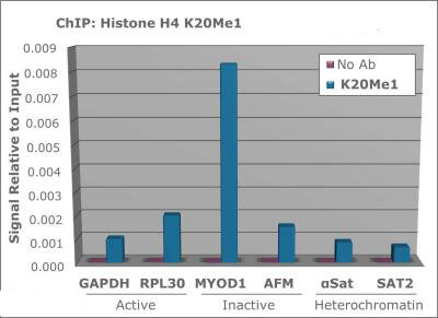
Chromatin Immunoprecipitation Rabbit Anti-Histone H4 [Monomethyl Lys20] Antibody. Chromatin from one million formaldehyde cross-linked Hela cells was used with 2ug of Anti-Histone H4K20me1 and 20ul of magnetic IgG beads per immunoprecipitation. A no antibody (No Ab) control was also used. Immunoprecipitated DNA was quantified using quantitative real-time PCR and SYBR green dye, then normalized to the non-precipitated input chromatin, which is equal to one.
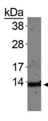
Western Blot of Rabbit Anti-Histone H4 [Monomethyl Lys20] Antibody. Lane 1: C. elegans embryo lysate. Load: 30 ug per lane. Primary antibody: Histone H4 [Monomethyl Lys20] at 1:500 for overnight at 4C. Secondary antibody: IRDye800TM rabbit secondary antibody at 1:10,000 for 45 min at RT. Block: 5% BLOTTO overnight at 4C. Predicted/Observed size: ~13 kDa. Other band(s): None.
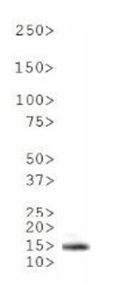
Western Blot of Rabbit Anti-Histone H4 [Monomethyl Lys20] Antibody. Lane 1: NIH-3T3 histone preps. Load: 30 ug per lane. Primary antibody: Histone H4 [Monomethyl Lys20] at 1:500 for overnight at 4C. Secondary antibody: IRDye800TM rabbit secondary antibody at 1:10,000 for 45 min at RT. Block: 5% BLOTTO overnight at 4C. Predicted/Observed size: ~13 kDa. Other band(s): None.
|
|
|
|
Immunofluorescence of Rabbit Anti Histone H4 [Monomethyl Lys20] Antibody. Tissue: HeLa cells. Fixation: 0.5% PFA. Antigen retrieval: Not required. Primary antibody: Histone H4 [Monomethyl Lys20] antibody at a 1:500 dilution for 1 h at RT. Secondary antibody: FITC secondary antibody at 1:10,000 for 45 min at RT. Localization: Histone H4 [Monomethyl Lys20] is nuclear and chromosomal. Staining: Histone H4 [Monomethyl Lys20] is expressed in green, nuclei and alpha tubulin are counterstained with DAPI (blue) and Dylight 550 (red).
|
|
|
| メーカー |
品番 |
包装 |
|
RKL
|
600-401-J01
|
50 UG
|
※表示価格について
| 当社在庫 |
なし
|
| 納期目安 |
約10日
|
| 保存温度 |
-20℃
|
|

