

| 別品名 |
H3.3B, H3 histone, family 3A, H3.3AH3F3H3F3B, histone H3.3, MGC87783, MGC87782, H3K9ac/K14ac
|
| 抗原部位 |
N-terminus
|
| 種由来 |
Human
|
| 標識物 |
Unlabeled
|
| 精製度 |
Affinity Purified
|
| 適用 |
Western Blot
Immuno Fluorescence
Dot Blot
|
| 免疫動物 |
Rabbit
|
| 抗体クラス |
IgG
|
| 交差種 |
Human
Mouse
Caenorhabditis elegans
|
| 翻訳後修飾 |
アセチル化
|
| GENE ID |
126961
|
| Accession No.(Gene/Protein) |
NP_001005464, Q71DI3
|
| Gene Symbol |
HIST2H3C
|
| 形状 |
滅菌済み液状品
|
| 参考文献 |
Campsteijn C., OvrebO JI., Karlsen BO., Thompson EM. (2012). Expansion of cyclin D and CDK1 paralogs in Oikopleura dioica, a chordate employing diverse cell cycle variants. Mol. Biol. Evol. 2012 Feb. 29:487-502. Yue C., Soboloff J., Gamero AM. (2012). Control of type I interferon-induced cell seath by Orai1-mediated calcium entry in T cells. J Biol Chem. 2012 Jan. 287:3207-3216. Kung VL., Khare S., Stehlik C., Bacon EM., Hughes AJ., Hauser AR. (2012). An rhs gene of Pseudomonas aeruginosa encodes a virulence protein that activates the inflammasome. Proc Natl Acad Sci USA. 2012 Jan. 109:1275-1280. Sullivan CP., Berg EA., Elliot-Bryant R., Fishman JB., Mckee AC., Morin PJ., et al. (2011). Pyroglutamate-Αβ 3 and 11 colocalize in amyloid plaques in Alzheimer's disease cerebral cortex with pyroglutamate-Αβ 11 forming the central core. Neurosci Lett. 2011 Nov 14. 505(2):109-12. Daftarian P., Kaifer AE., Li W., Blomberg BB., Frasca D., Roth F., et al. (2011). Peptide-conjugated PAMAM dendrimer as a universal platform for antigen presenting cell targeting and effective DNA-based vaccinations. Cancer Res. 2011 Oct 10. 71(24):7452-62.
|
| [注意事項] |
濃度はロットによって異なる可能性があります。メーカーDS及びCoAからご確認ください。
|
|
※サムネイル画像をクリックすると拡大画像が表示されます。
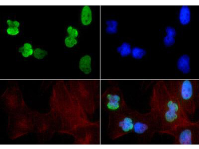
Immunofluorescence of Rabbit Anti Histone H3 [ac Lys9, ac Lys14] Antibody. Tissue: HeLa cells. Fixation: 0.5% PFA. Antigen retrieval: Not required. Primary antibody: Histone H3 [ac Lys9, ac Lys14] antibody at a 1:200 dilution for 1 h at RT. Secondary antibody: FITC secondary antibody at 1:10,000 for 45 min at RT. Localization: Histone H3 [ac Lys9, ac Lys14] is nuclear and chromosomal. Staining: Histone H3 [ac Lys9, ac Lys14] is expressed in green, nuclei and actin are counterstained with Dapi (blue) and Phalloidin (red).
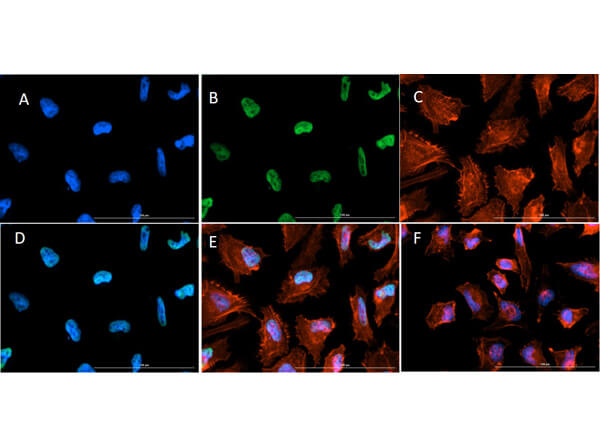
ImmunoFluorescence of Rabbit Anti-Histone H3 K9ac/K14ac Antibody. Cells: HeLa Cells. Fixation: 4% PFA. Permeabilization: 0.3% Triton X-100. Primary Antibody: Anti-Histone H3 [ac Lys9, ac Lys14] at 5ug/mL overnight at 2-8C. Secondary Antibody: Goat Anti-Rabbit IgG DyLightTM488 (p/n 611-141-122) at 5ug/mL for 1hr at RT. Nuclear Counterstain: DAPI. Actin Filament Stain: Texas Red - X Phallodin. Staining: (A) DAPI. (B) K9 AC/K14 AC + DyLightTM488. (C) Actin-X Phallodin. (D) Merge A+B. (E) Merge A+B+C. (F) Secondary Only Merge. Expected Location: Nuclear and Chromosomal.
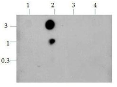
Dot Blot of Rabbit Histone H3 [K9ac, K14ac] Antibody. Lane 1: K9-K14. Lane 2: K9-KAc-K14-Kac. Lane 3: K9-K14-Kac. Lane 4: K9-Ac-K14. Load: 0.3, 1, and 3 μg of peptide. Primary antibody: Histone H3 [ac Lys9, ac Lys14] antibody for 45 min at 4°C. Secondary antibody: HRP rabbit secondary antibody at 1:10,000 for 45 min at RT. Block: 5% BLOTTO A overnight at 4°C.
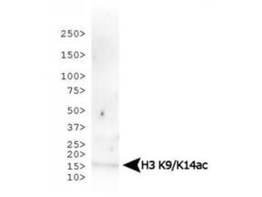
Western Blot of Rabbit Anti-Histone H3 [ac Lys9, ac Lys14] Antibody. Lane 1: C. elegans embryo lysate. Load: 30 ug per lane. Primary antibody: Histone H3 [ac Lys9, ac Lys14] at 1:500 for overnight at 4C. Secondary antibody: IRDye800TM rabbit secondary antibody at 1:10,000 for 45 min at RT. Block: 5% BLOTTO overnight at 4C. Predicted/Observed size: ~15 kDa. Other band(s): None.

Western Blot of Rabbit Anti-Histone H3 [ac Lys9, ac Lys14] Antibody. Lane 1: NIH-3T3 histone preps. Load: 30 ug per lane. Primary antibody: Histone H3 [ac Lys9, ac Lys14] at 1:500 for overnight at 4C. Secondary antibody: IRDye800TM rabbit secondary antibody at 1:10,000 for 45 min at RT. Block: 5% BLOTTO overnight at 4C. Predicted/Observed size: ~15 kDa. Other band(s): None.
|
|
|
|
Immunofluorescence of Rabbit Anti Histone H3 [ac Lys9, ac Lys14] Antibody. Tissue: HeLa cells. Fixation: 0.5% PFA. Antigen retrieval: Not required. Primary antibody: Histone H3 [ac Lys9, ac Lys14] antibody at a 1:200 dilution for 1 h at RT. Secondary antibody: FITC secondary antibody at 1:10,000 for 45 min at RT. Localization: Histone H3 [ac Lys9, ac Lys14] is nuclear and chromosomal. Staining: Histone H3 [ac Lys9, ac Lys14] is expressed in green, nuclei and actin are counterstained with Dapi (blue) and Phalloidin (red).
|
|
|
| メーカー |
品番 |
包装 |
|
RKL
|
600-401-I73
|
50 UG
|
※表示価格について
| 当社在庫 |
なし
|
| 納期目安 |
約10日
|
| 保存温度 |
-20℃
|
|

