|
※サムネイル画像をクリックすると拡大画像が表示されます。
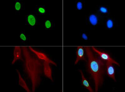
Immunofluorescence of Rabbit Anti-Histone H3 [ac Lys9] Antibody. Tissue: HeLa cells. Fixation: 0.5% PFA. Antigen retrieval: Not required. Primary antibody: Histone H3 [ac Lys9] antibody at a 1:100 dilution for 1 h at RT. Secondary antibody: FITC secondary antibody at 1:10,000 for 45 min at RT. Localization: Histone H3 [ac Lys9] is nuclear and chromosomal. Staining: Histone H3 [ac Lys9] is expressed in green.
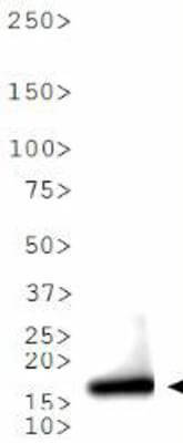
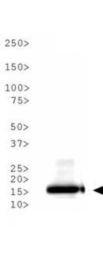
Western Blot of Rabbit Anti-Histone H3 [ac Lys9] Antibody. Lane 1: HeLa histone preps. Load: 30 μg per lane. Primary antibody: Histone H3 [ac Lys9] at 1:500 for overnight at 4°C. Secondary antibody: IRDye800? rabbit secondary antibody at 1:10,000 for 45 min at RT. Block: 5% BLOTTO overnight at 4°C. Predicted/Observed size: ~15 kDa. Other band(s): None.
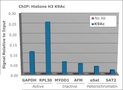
Chromatin Immunoprecipitation of Rabbit Anti-Histone H3 [ac Lys9] Antibody. Chromatin from one million formaldehyde cross-linked Hela cells was used with 2 ug of Anti-Histone H3 K9ac was used to IP DNA from fixed Hela cells alongside a no antibody (No Ab) control. DNA was measured by qRT-PCR and normalized to total input (input=1).
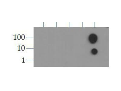
Dot Blot of Rabbit Histone H3 [ac Lys9] Antibody. Lane 1: K9. Lane 2: K9 Me1. Lane 3: K9 Me2. Lane 4: K9 Me3. Lane 5: K9ac. Load: 1, 10, and 100 picomoles of peptide. Primary antibody: Histone H3 [ac Lys9] antibody at 1:1000 for 45 min at 4°C. Secondary antibody: Dylight?488 rabbit secondary antibody at 1:10,000 for 45 min at RT. Block: 5% BLOTTO overnight at 4°C.
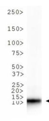
Western Blot of Rabbit Anti-Histone H3 [ac Lys9] Antibody. Lane 1: NIH-3T3 histone preps. Load: 30 μg per lane. Primary antibody: Histone H3 [ac Lys9] at 1:500 for overnight at 4°C. Secondary antibody: IRDye800? rabbit secondary antibody at 1:10,000 for 45 min at RT. Block: 5% BLOTTO overnight at 4°C. Predicted/Observed size: ~15 kDa. Other band(s): None.
Western Blot of Rabbit Anti-Histone H3 [ac Lys9] Antibody. Lane 1: C. elegans embryo lysate. Load: 30 μg per lane. Primary antibody: Histone H3 [ac Lys9] at 1:500 for overnight at 4°C. Secondary antibody: IRDye800? rabbit secondary antibody at 1:10,000 for 45 min at RT. Block: 5% BLOTTO overnight at 4°C. Predicted/Observed size: ~15 kDa. Other band(s): None.
|
|
|
|
Immunofluorescence of Rabbit Anti-Histone H3 [ac Lys9] Antibody. Tissue: HeLa cells. Fixation: 0.5% PFA. Antigen retrieval: Not required. Primary antibody: Histone H3 [ac Lys9] antibody at a 1:100 dilution for 1 h at RT. Secondary antibody: FITC secondary antibody at 1:10,000 for 45 min at RT. Localization: Histone H3 [ac Lys9] is nuclear and chromosomal. Staining: Histone H3 [ac Lys9] is expressed in green.
|
|
| 別品名 |
rabbit anti-Histone H3 Ac Lys9 antibody, H3.3B, H3 histone, family 3A, H3.3AH3F3H3F3B, histone H3.3, MGC87783, MGC87782, H3K9ac
|
| 交差種 |
Human
Mouse
Caenorhabditis elegans
|
| 適用 |
Western Blot
Immuno Fluorescence
Chromatin Immunoprecipitation
Dot Blot
|
| 免疫動物 |
Rabbit
|
| 抗原部位 |
N-terminus
|
| 標識物 |
Unlabeled
|
| 精製度 |
Affinity Purified
|
| 翻訳後修飾 |
アセチル化
|
| GENE ID |
126961
|
| Accession No.(Gene/Protein) |
NP_001005464, Q71DI3
|
| Gene Symbol |
HIST2H3C
|
| 参考文献 |
[Pub Med ID]29123096
|
| [注意事項] |
濃度はロットによって異なる可能性があります。メーカーDS及びCoAからご確認ください。
|
|
| メーカー |
品番 |
包装 |
|
RKL
|
600-401-I72
|
50 UG
|
※表示価格について
| 当社在庫 |
なし
|
| 納期目安 |
約10日
|
| 保存温度 |
-20℃
|
|
※当社では商品情報の適切な管理に努めておりますが、表示される法規制情報は最新でない可能性があります。
また法規制情報の表示が無いものは、必ずしも法規制に非該当であることを示すものではありません。
商品のお届け前に最新の製品法規制情報をお求めの際はこちらへお問い合わせください。
|
※当社取り扱いの試薬・機器製品および受託サービス・創薬支援サービス(納品物、解析データ等)は、研究用としてのみ販売しております。
人や動物の医療用・臨床診断用・食品用としては、使用しないように、十分ご注意ください。
法規制欄に体外診断用医薬品と記載のものは除きます。
|
|
※リンク先での文献等のダウンロードに際しましては、掲載元の規約遵守をお願いします。
|
|
※CAS Registry Numbers have not been verified by CAS and may be inaccurate.
|
