|
※サムネイル画像をクリックすると拡大画像が表示されます。
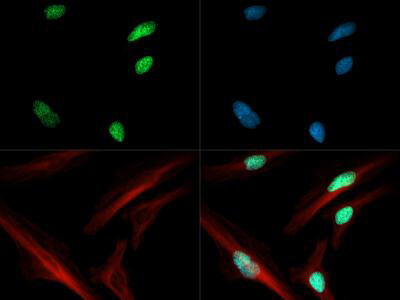
Immunofluorescence of Rabbit Anti-Histone H3 [ac Lys4] Antibody. Tissue: HeLa cells. Fixation: 0.5% PFA. Antigen retrieval: Not required. Primary antibody: Histone H3 [ac Lys4] antibody at a 1:50 dilution for 1 h at RT. Secondary antibody: Dylight 488 secondary antibody at 1:10,000 for 45 min at RT. Localization: Histone H3 [ac Lys4] is nuclear and chromosomal. Staining: Histone H3 [ac Lys4] is expressed in green and the nuclei and alpha-tubulin are counterstained with DAPI (blue) and Dylight 566 (red).
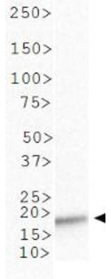
Western Blot of Rabbit Anti-Histone H3 [ac Lys4] Antibody. Lane 1: C. elegans embryo lysate. Load: 30 μg per lane. Primary antibody: Histone H3 [ac Lys4] at 1:500 for overnight at 4°C. Secondary antibody: IRDye800? rabbit secondary antibody at 1:10,000 for 45 min at RT. Block: 5% BLOTTO overnight at 4°C. Predicted/Observed size: ~15-16 kDa. Other band(s): None.
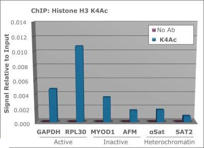
Chromatin Immunoprecipitation of Rabbit Anti-Histone H3 [ac Lys4] Antibody. Chromatin from one million formaldehyde cross-linked Hela cells was used with 2ug of Anti-Histone H3 K4ac and 20ul of magnetic IgG beads per immunoprecipitation. A no antibody (No Ab) control was also used. Immunoprecipitated DNA was quantified using quantitative real-time PCR and SYBR green dye, then normalized to the non-precipitated input chromatin, which is equal to one.
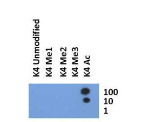
Dot Blot of Rabbit Histone H3 [ac Lys4] Antibody. Lane 1: K4 umodified. Lane 2: K4 Me1. Lane 3: K4 Me2. Lane 4: K4 Me3. Lane 5: K4 Ac. Load: 1, 10, and 100 picomoles of peptide. Primary antibody: Histone H3 [ac Lys4] antibody at 1:1000 for 45 min at 4°C. Secondary antibody: Dylight?488 rabbit secondary antibody at 1:10,000 for 45 min at RT. Block: 5% BLOTTO overnight at 4°C.
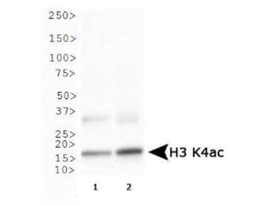
Western Blot of Rabbit Anti-Histone H3 [ac Lys4] Antibody. Lane 1: HeLa histone prep. Lane 2: NIH-3T3 prep lysates. Load: 30 μg per lane. Primary antibody: Histone H3 [ac Lys4] at 1:500 for overnight at 4°C. Secondary antibody: IRDye800? rabbit secondary antibody at 1:10,000 for 45 min at RT. Block: 5% BLOTTO overnight at 4°C. Predicted/Observed size: ~15 kDa. Other band(s): None.
|
|
|
|
Immunofluorescence of Rabbit Anti-Histone H3 [ac Lys4] Antibody. Tissue: HeLa cells. Fixation: 0.5% PFA. Antigen retrieval: Not required. Primary antibody: Histone H3 [ac Lys4] antibody at a 1:50 dilution for 1 h at RT. Secondary antibody: Dylight 488 secondary antibody at 1:10,000 for 45 min at RT. Localization: Histone H3 [ac Lys4] is nuclear and chromosomal. Staining: Histone H3 [ac Lys4] is expressed in green and the nuclei and alpha-tubulin are counterstained with DAPI (blue) and Dylight 566 (red).
|
|
| 別品名 |
rabbit anti-Histone H3 Ac Lys4 antibody, H3.3AH3F3H3F3B, H3.3B, H3 histone, family 3A, histone H3.3, MGC87783, MGC87782, H3K4ac
|
| 交差種 |
Human
Mouse
Caenorhabditis elegans
|
| 適用 |
Western Blot
Immuno Fluorescence
Chromatin Immunoprecipitation
Dot Blot
|
| 免疫動物 |
Rabbit
|
| 抗原部位 |
N-terminus
|
| 標識物 |
Unlabeled
|
| 精製度 |
Affinity Purified
|
| 翻訳後修飾 |
アセチル化
|
| GENE ID |
126961
|
| Accession No.(Gene/Protein) |
NP_001005464, Q71DI3
|
| Gene Symbol |
HIST2H3C
|
| 参考文献 |
Campsteijn C., OvrebO JI., Karlsen BO., Thompson EM. (2012). Expansion of cyclin D and CDK1 paralogs in Oikopleura dioica, a chordate employing diverse cell cycle variants. Mol. Biol. Evol. 2012 Feb. 29:487-502. Yue C., Soboloff J., Gamero AM. (2012). Control of type I interferon-induced cell seath by Orai1-mediated calcium entry in T cells. J Biol Chem. 2012 Jan. 287:3207-3216. Kung VL., Khare S., Stehlik C., Bacon EM., Hughes AJ., Hauser AR. (2012). An rhs gene of Pseudomonas aeruginosa encodes a virulence protein that activates the inflammasome. Proc Natl Acad Sci USA. 2012 Jan. 109:1275-1280. Sullivan CP., Berg EA., Elliot-Bryant R., Fishman JB., Mckee AC., Morin PJ., et al. (2011). Pyroglutamate-Αβ 3 and 11 colocalize in amyloid plaques in Alzheimer's disease cerebral cortex with pyroglutamate-Αβ 11 forming the central core. Neurosci Lett. 2011 Nov 14. 505(2):109-12. Daftarian P., Kaifer AE., Li W., Blomberg BB., Frasca D., Roth F., et al. (2011). Peptide-conjugated PAMAM dendrimer as a universal platform for antigen presenting cell targeting and effective DNA-based vaccinations. Cancer Res. 2011 Oct 10. 71(24):7452-62.
|
|
| メーカー |
品番 |
包装 |
|
RKL
|
600-401-I60
|
50 UG
|
※表示価格について
| 当社在庫 |
なし
|
| 納期目安 |
約10日
|
| 保存温度 |
-20℃
|
|
※当社では商品情報の適切な管理に努めておりますが、表示される法規制情報は最新でない可能性があります。
また法規制情報の表示が無いものは、必ずしも法規制に非該当であることを示すものではありません。
商品のお届け前に最新の製品法規制情報をお求めの際はこちらへお問い合わせください。
|
※当社取り扱いの試薬・機器製品および受託サービス・創薬支援サービス(納品物、解析データ等)は、研究用としてのみ販売しております。
人や動物の医療用・臨床診断用・食品用としては、使用しないように、十分ご注意ください。
法規制欄に体外診断用医薬品と記載のものは除きます。
|
|
※リンク先での文献等のダウンロードに際しましては、掲載元の規約遵守をお願いします。
|
|
※CAS Registry Numbers have not been verified by CAS and may be inaccurate.
|
