|
※サムネイル画像をクリックすると拡大画像が表示されます。
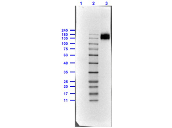
Western Blot of Rabbit TrueBlotR ULTRA: Anti-Rabbit IgG HRP. Lane 1: Rabbit IgG WM -reduced (p/n 011-0102) [0.1μg]. Lane 2: Opal Prestained Molecular Weight Marker (p/n MB-210-0500). Lane 3: Rabbit IgG WM non-reduced (p/n 011-0102) [0.1μg]. Antibody: Rabbit TrueBlotR ULTRA: Anti-Rabbit IgG HRP at 1.0μg/mL overnight at 4°C. Blocking Buffer for Fluorescent Western Blotting (p/n MB-070) for 60mins at RT. Expect: recognizes the Rabbit IgG, only under non-reducing condition. Exposure: 0.45sec.
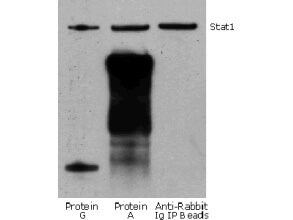
Rabbit TrueBlotR IP / Western Blot: Jurkat cell lysate (0.5 ml of 1x10e7 cells/ml) was incubated with rabbit anti-human Stat1 and immunoprecipitated using Protein G, Protein A and Anti-Rabbit Ig IP Beads. Precipitate from 5x10e5 cells was subjected to electrophoresis, transferred to a PVDF membrane, and Western blotted with anti-Stat1 using Rabbit TrueBlotR ULTRA: Anti-Rabbit IgG HRP
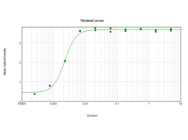
ELISA results of Rabbit TrueBlotR ULTRA: Anti-Rabbit IgG HRP tested against purified Rabbit IgG protein. Each well was coated in duplicate with 1.0 μg of Rabbit IgG (p/n 011-0102). The starting dilution of antibody was 5μg/ml and the X-axis represents the Log10 of a 3-fold dilution. The titer is 1:450,000. This titration is a 4-parameter curve fit where the IC50 is defined as the titer of the antibody. Assay performed using 3% fish gelatin as blocking buffer and TMB substrate p/n TMBE-1000.
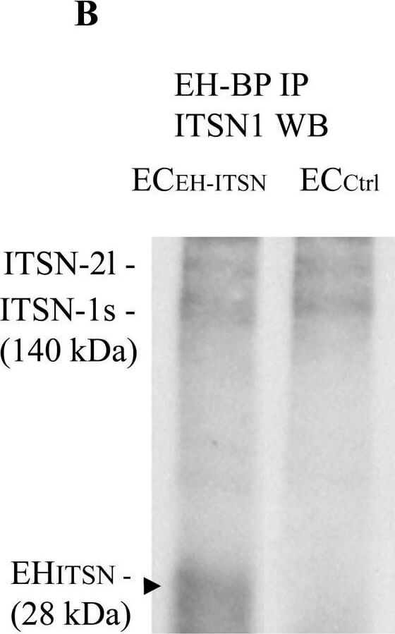
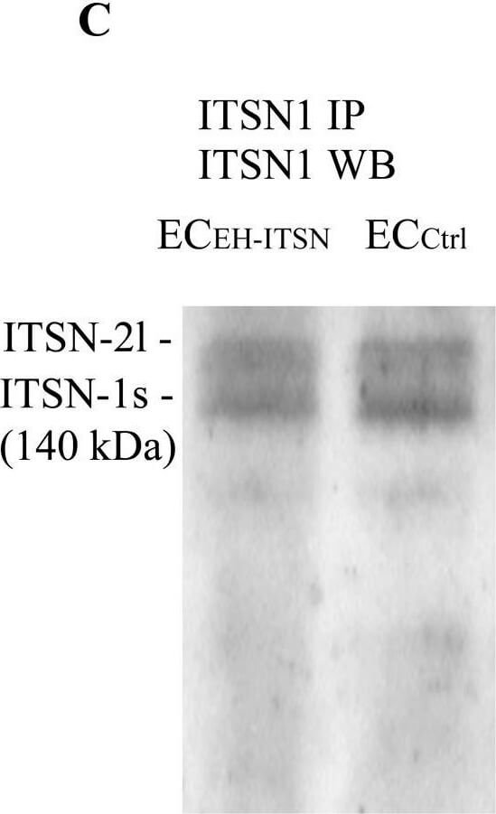
Intersectin-1s (ITSN) interacts via the EH domains with the EHBP1. ECs lysates (250 μg total protein) were subjected to immunoprecipitation with anti-EHBP1 Ab (1 μg), followed by WB with EHBP1 (A) and ITSN1 (B) Abs. EHBP1 Ab brings down the EHBP1 protein as well as ITSN. The 55 kDa immunoreactivity in panel A is cross-reactivity with the IgG heavy chain. The EHBP1 Ab immunoprecipitates the Myc-EHITSN from the stable transfected ECEH-ITSN lysates (B, arrowhead). (C). ECs lysates (250 μg total protein) were subjected to immunoprecipitation with anti-ITSN1 Ab (1 μg), followed by WB with ITSN1 Ab. ITSN1 Ab brings down ITSN protein in both ECEH-ITSN and ECCtrl lysates. The upper ITSN immunoreactivity (190 kDa), belongs to the ITSN-2 long isoform (ITSN-2l). For immunoprecipitation studies (B,C), the rabbit IgG TrueBlot Ab HRP ULTRA conjugated which enables detection of immunoblotted target protein bands, without interfering with the immunoprecipitating IgG heavy and light chains has been used. (D) Densitometric analysis of immunoprecipitated ITSN in both ECEH-ITSN and ECCtrl lysates. Data are expressed as ratio of ITSN immunoprecipitated by EHBP1 Ab / ITSN immunoprecipitated by ITSN Ab (D). ?p < 0.05. (E,F). Double anti-ITSN Ab anti-rabbit IgG Alexa Fluor 594-conjugated (E) / anti-EHBP1 Ab ? anti mouse IgG Alexa Fluor 488-conjugated (F). The merged image reveals significant co-localization ITSN/EHBP1, both in the cytosol and at the plasma membrane (G). (H) The magnification of the boxed area in G, highlights the significant co-localization ITSN/EHBP1 at the plasma membrane level (arrows) and cytosol (arrowheads). Bars: 10 μm (E?G); 5 μm (H); n = 5. Figure provided by CiteAb. Source: Front Physiol, PMID: 30333761.
Intersectin-1s (ITSN) interacts via the EH domains with the EHBP1. ECs lysates (250 μg total protein) were subjected to immunoprecipitation with anti-EHBP1 Ab (1 μg), followed by WB with EHBP1 (A) and ITSN1 (B) Abs. EHBP1 Ab brings down the EHBP1 protein as well as ITSN. The 55 kDa immunoreactivity in panel A is cross-reactivity with the IgG heavy chain. The EHBP1 Ab immunoprecipitates the Myc-EHITSN from the stable transfected ECEH-ITSN lysates (B, arrowhead). (C). ECs lysates (250 μg total protein) were subjected to immunoprecipitation with anti-ITSN1 Ab (1 μg), followed by WB with ITSN1 Ab. ITSN1 Ab brings down ITSN protein in both ECEH-ITSN and ECCtrl lysates. The upper ITSN immunoreactivity (190 kDa), belongs to the ITSN-2 long isoform (ITSN-2l). For immunoprecipitation studies (B,C), the rabbit IgG TrueBlot Ab HRP ULTRA conjugated which enables detection of immunoblotted target protein bands, without interfering with the immunoprecipitating IgG heavy and light chains has been used. (D) Densitometric analysis of immunoprecipitated ITSN in both ECEH-ITSN and ECCtrl lysates. Data are expressed as ratio of ITSN immunoprecipitated by EHBP1 Ab / ITSN immunoprecipitated by ITSN Ab (D). ?p < 0.05. (E,F). Double anti-ITSN Ab anti-rabbit IgG Alexa Fluor 594-conjugated (E) / anti-EHBP1 Ab ? anti mouse IgG Alexa Fluor 488-conjugated (F). The merged image reveals significant co-localization ITSN/EHBP1, both in the cytosol and at the plasma membrane (G). (H) The magnification of the boxed area in G, highlights the significant co-localization ITSN/EHBP1 at the plasma membrane level (arrows) and cytosol (arrowheads). Bars: 10 μm (E?G); 5 μm (H); n = 5. Figure provided by CiteAb. Source: Front Physiol, PMID: 30333761.
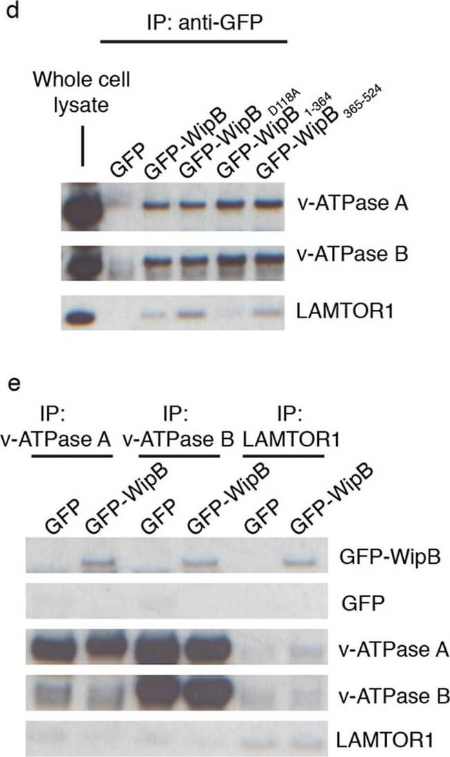
WipB is targeted to lysosomes by its C-terminal domain where it interacts with components of the lysosomal nutrient sensing system (a) HeLa cells expressing GFP-fusion proteins of WipB, WipBD118A, WipB1-364 and WipB365-524. Scale bar, 10?μm. (b) HeLa cells expressing GFP-WipB (green) stained using an anti-LAMP-1 antibody (Magenta) after permeabilisation and fixation. Scale bar, 10?μm. (c) HeLa cells expressing GFP-WipB (green) were incubated with Lysotracker (red) for 15?min before fixation. Scale bar, 10?μm. (d) SDS-PAGE of fractions following co-immunoprecipitation of GFP, GFP-WipB or the indicated GFP-WipB derivatives from HeLa cell lysates using anti-GFP antibody and immunoblotting with anti-v-ATPase A, -v-ATPase B or LAMTOR1 antibodies. A cropped blot is here displayed and the corresponding full-length blot is included in the supplementary information. (e) SDS-PAGE of fractions following co-immunoprecipitation of v-ATPase A, v-ATPase B or LAMTOR1 from lysates of HeLa cells expressing GFP or GFP-WipB and immunoblotting with anti-GFP antibody. A cropped blot is here displayed and the corresponding full-length blot is included in the supplementary information. Figure provided by CiteAb. Source: Sci Rep, PMID: 28842705.
|
|
|
|
Western Blot of Rabbit TrueBlotR ULTRA: Anti-Rabbit IgG HRP. Lane 1: Rabbit IgG WM -reduced (p/n 011-0102) [0.1μg]. Lane 2: Opal Prestained Molecular Weight Marker (p/n MB-210-0500). Lane 3: Rabbit IgG WM non-reduced (p/n 011-0102) [0.1μg]. Antibody: Rabbit TrueBlotR ULTRA: Anti-Rabbit IgG HRP at 1.0μg/mL overnight at 4°C. Blocking Buffer for Fluorescent Western Blotting (p/n MB-070) for 60mins at RT. Expect: recognizes the Rabbit IgG, only under non-reducing condition. Exposure: 0.45sec.
|
|
| 別品名 |
Anti-Rabbit IgG HRP, TrueBlot, HRP TrueBlot ULTRA, Peroxidase TrueBlot, TrueBlot for IP/WB, TrueBlot for immunoprecipitation, TrueBlot for western blotting
|
| 交差種 |
Rabbit
|
| 適用 |
Western Blot
Enzyme Linked Immunosorbent Assay
Immunoprecipitation
|
| 免疫動物 |
Mouse
|
| クローン |
eB182
|
| 標識物 |
Horseradish Peroxidase
|
| 精製度 |
Affinity Purified
|
| 構成内容 |
ミニゲルで約80ブロット分の試薬が含まれます。
|
| 参考文献 |
[Pub Med ID]18949366
|
| [注意事項] |
濃度はロットによって異なる可能性があります。メーカーDS及びCoAからご確認ください。
|
|
| メーカー |
品番 |
包装 |
|
RKL
|
18-8816-33
|
200 UL
|
※表示価格について
|
※当社では商品情報の適切な管理に努めておりますが、表示される法規制情報は最新でない可能性があります。
また法規制情報の表示が無いものは、必ずしも法規制に非該当であることを示すものではありません。
商品のお届け前に最新の製品法規制情報をお求めの際はこちらへお問い合わせください。
|
※当社取り扱いの試薬・機器製品および受託サービス・創薬支援サービス(納品物、解析データ等)は、研究用としてのみ販売しております。
人や動物の医療用・臨床診断用・食品用としては、使用しないように、十分ご注意ください。
法規制欄に体外診断用医薬品と記載のものは除きます。
|
|
※リンク先での文献等のダウンロードに際しましては、掲載元の規約遵守をお願いします。
|
|
※CAS Registry Numbers have not been verified by CAS and may be inaccurate.
|
