
| 別品名 |
DLG4, SAP90, Synapse-associated protein 90, Postsynaptic density protein 95, Disks large homolog 4
|
| 種由来 |
Rat
|
| 標識物 |
Unlabeled
|
| 精製度 |
Ig fraction - Protein G
|
| 適用 |
Western Blot
Immunohistochemistry
Immuno Fluorescence
|
| 免疫動物 |
Mouse
|
| 抗体クラス |
IgG2a
|
| クローン |
7E3
|
| 交差種 |
Mouse
Rat
Bovine
|
| GENE ID |
13385
|
| Accession No.(Gene/Protein) |
NP_031890, P31016
|
| Gene Symbol |
Dlg4
|
| 形状 |
滅菌済み液状品
|
| 参考文献 |
1. Chetkovich D.M., Bunn R.C., Kuo S.H., Kawasaki Y., Kohwi M., and Bredt D.S. (2002) J Neurosci. 22(15): 6415-25. 2. Cao J., Viholainen J.I., Dart C., Warwick H.K., Levland M.L. and Courtney M.J. (2005) J Cell Biol. 168(1): 117-26. 3. Kennedy M. (1997) Trends in Neurosci. 6: 264-268. 4. Irie M. et al. (1997) Science 277(5331): 1511-5. 5. Cai C. et al. (2006) J Biol Chem. 281: 4267-73. 6. Yao W.D. et al. (2004) Neuron 41: 625-38. 7. Cline H. (2005) Curr Biol. 15: R203-5.
|
| [注意事項] |
濃度はロットによって異なる可能性があります。メーカーDS及びCoAからご確認ください。
|
|
※サムネイル画像をクリックすると拡大画像が表示されます。
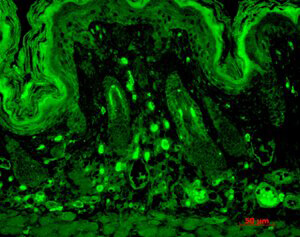
Immunohistochemistry of mouse anti Slo2.1 antibody. Tissue: Sections of mouse backskin. Primary Antibody: Slo2.1 antibody at 1 μg/mL for 1h at RT. Secondary antibody: Peroxidase mouse secondary at 1:10,000 for 45 min at RT. Localization: Membrane. Staining: Slo2.1 as white signal.
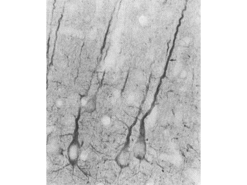
Immunocytochemistry of Mouse anti-PSD95 antibody. Tissue: Rat neocortex. Fixation: N/A Antigen retrieval: not required. Primary antibody: PSD95 antibody at 10 ug/mL for 1h at RT. Secondary antibody: Fluorescein mouse secondary antibody at 1:10,000 for 45 min at RT. Localization: PSD95 is cell membrane and cell junctions. Staining: PSD95 as precipitated black signal.
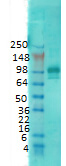
Western Blot of mouse anti-PSD95 total antibody. Lane 1: Rat Membrane. Primary antibody: PSD95 total antibody at 1:1000 for overnight at 4C. Secondary antibody: Goat anti-mouse IgG HRP secondary antibody at 1:10,000 for 45 min at RT. Block: 5% Blotto overnight 4C. Predicted/Observed size: 80.4 kDa/100kD. Other band(s): ~80kDa and ~50kDa in rat and mouse samples.
|
|
|
|
Immunohistochemistry of mouse anti Slo2.1 antibody. Tissue: Sections of mouse backskin. Primary Antibody: Slo2.1 antibody at 1 μg/mL for 1h at RT. Secondary antibody: Peroxidase mouse secondary at 1:10,000 for 45 min at RT. Localization: Membrane. Staining: Slo2.1 as white signal.
|
|
|
| メーカー |
品番 |
包装 |
|
RKL
|
200-301-G33
|
100 UG
|
※表示価格について
| 当社在庫 |
なし
|
| 納期目安 |
約10日
|
| 保存温度 |
-20℃
|
|

