|
※サムネイル画像をクリックすると拡大画像が表示されます。
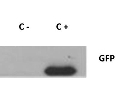
Anti-GFP Antibody - Western Blot. Western blot of GFP protein detected with polyclonal anti-GFP antibody. Wild type GFP (0.1 ug) was used to spike 30 ug of a HeLa whole cell lysate. This antibody detects a 27 kD band corresponding to the epitope tag GFP. A 4-20% Tris-Glycine gradient gel was used for SDS-PAGE. The protein was transferred to nitrocellulose using standard methods. After blocking with 5% BLOTTO in PBS, the membrane was probed overnight at 4C with the primary antibody diluted in 5% BLOTTO to 1:1000, followed by washes and reaction with a 1:10000 dilution of IRDye 800 conjugated Goat-a-Rabbit IgG [H&L] MX10 (. IRDye 800 fluorescence image was captured using the Odyssey Infrared Imaging System developed by LI-COR. IRDye is a trademark of LI-COR, Inc. Other detection systems will yield similar results.
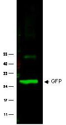
Western blot of Rabbit anti-GFP antibody. Lane 1: Wild type GFP (0.1 g) was used to spike HeLa whole cell lysate. Lane 2: none. Load: 30 ug per lane. Primary antibody: GFP antibody at 1:1000 for overnight at 4C. Secondary antibody: IRDye800 Goat-a-Rabbit IgG [H&L] MX10 (611-132-122) at 1:10000 for 45 min at RT. Block: 5% BLOTTO in PBS overnight at 4C. Predicted/Observed size: 27 kDa for epitope tag GFP. Other band(s): none.
Immuno-microscopy of Rabbit anti-GFP antibody. Monocyte derived dendritic cells and dermal macrophages were challenged and directly visualized with eGFP labeled Dengue virus to localize sequestration of virus particles in the different cells (upper). The location of the GFP was confirmed by TEM (lower magnified view) using rabbit anti GFP Primary antibody (1:200) and a gold labeled secondary antibody. As referenced in: Kwan W-H, Navarro-Sanchez E, Dumortier H, Decossas M, Vachon H, et al. (2008) Dermal-Type Macrophages Expressing CD209/DC-SIGN Show Inherent Resistance to Dengue Virus Growth. PLoS Negl Trop Dis 2(10): e311. doi:10.1371/journal.pntd.0000311

|
|
|
|
Anti-GFP Antibody - Western Blot. Western blot of GFP protein detected with polyclonal anti-GFP antibody. Wild type GFP (0.1 ug) was used to spike 30 ug of a HeLa whole cell lysate. This antibody detects a 27 kD band corresponding to the epitope tag GFP. A 4-20% Tris-Glycine gradient gel was used for SDS-PAGE. The protein was transferred to nitrocellulose using standard methods. After blocking with 5% BLOTTO in PBS, the membrane was probed overnight at 4C with the primary antibody diluted in 5% BLOTTO to 1:1000, followed by washes and reaction with a 1:10000 dilution of IRDye 800 conjugated Goat-a-Rabbit IgG [H&L] MX10 (. IRDye 800 fluorescence image was captured using the Odyssey Infrared Imaging System developed by LI-COR. IRDye is a trademark of LI-COR, Inc. Other detection systems will yield similar results.
|
|
| 別品名 |
GFP
|
| 種由来 |
Aequorea victoria
|
| 交差種 |
Aequorea victoria
|
| 適用 |
Western Blot
Enzyme Linked Immunosorbent Assay
Immunohistochemistry
Immuno Fluorescence
|
| 免疫動物 |
Rabbit
|
| 抗体クラス |
IgG
|
| 標識物 |
Unlabeled
|
| 精製度 |
Affinity Purified
|
| Gene Symbol |
GFP
|
| 使用文献 |
[Pub Med ID]21127067, 21278252, 21792173, 22322860, 22511115
|
|
| メーカー |
品番 |
包装 |
|
LSP
|
LS-C154219-100
|
100 UG
|
※表示価格について
| 当社在庫 |
なし
|
| 納期目安 |
約1ヶ月
|
| 保存温度 |
-20℃
|
|
※当社では商品情報の適切な管理に努めておりますが、表示される法規制情報は最新でない可能性があります。
また法規制情報の表示が無いものは、必ずしも法規制に非該当であることを示すものではありません。
商品のお届け前に最新の製品法規制情報をお求めの際はこちらへお問い合わせください。
|
※当社取り扱いの試薬・機器製品および受託サービス・創薬支援サービス(納品物、解析データ等)は、研究用としてのみ販売しております。
人や動物の医療用・臨床診断用・食品用としては、使用しないように、十分ご注意ください。
法規制欄に体外診断用医薬品と記載のものは除きます。
|
|
※リンク先での文献等のダウンロードに際しましては、掲載元の規約遵守をお願いします。
|
