|
※サムネイル画像をクリックすると拡大画像が表示されます。
Blocking Buffer for Fluorescent Western Blotting
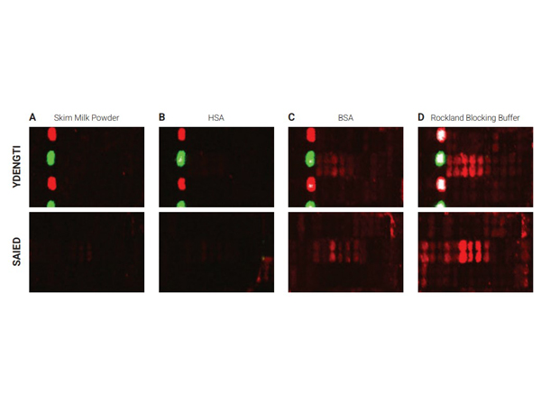
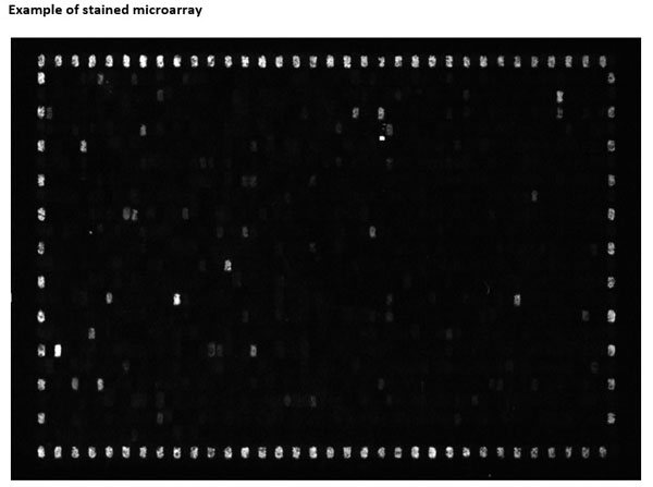
Selected sections of the PEPperCHIPR Peptide Microarrays after assay with different blocking reagents. The microarrays were blocked for 30 minutes with either 2% skim milk powder (A), 1% HSA (B), 1% BSA (C) or 100% Rockland Blocking Buffer [p/n MB-070] (D), respectively. A human serum sample was assayed at dilution 1:200, followed by detection with secondary goat Anti-Human IgG (H+L) DyLight? 680 Antibody [p/n 609-144-123]. Red spots = sample responses and polio control peptides, green spots = HA control peptides. The underlying binding motifs of the respective sections are indicated on the left.
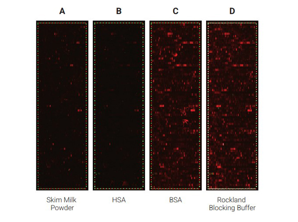
Comparison of the performance of different blocking reagents in epitope mappings with PEPperCHIPR Peptide Microarrays.The PEPperCHIPR Peptide Microarrays were blocked for 30 minutes with either 2% skim milk powder (A), 1% HSA (B), 1% BSA (C) or 100% Rockland Blocking Buffer [p/n MB-070] (D). A human serum sample was assayed at dilution 1:200, followed by detection with secondary goat anti-Human IgG (H+L) DyLight? 680 Antibody [p/n 609-144-123] and a control anti-HA (12CA5)-DyLight? 800 Antibody. Red spots = sample IgG response and frame of polio control peptides, green spots = frame of HA control peptides.
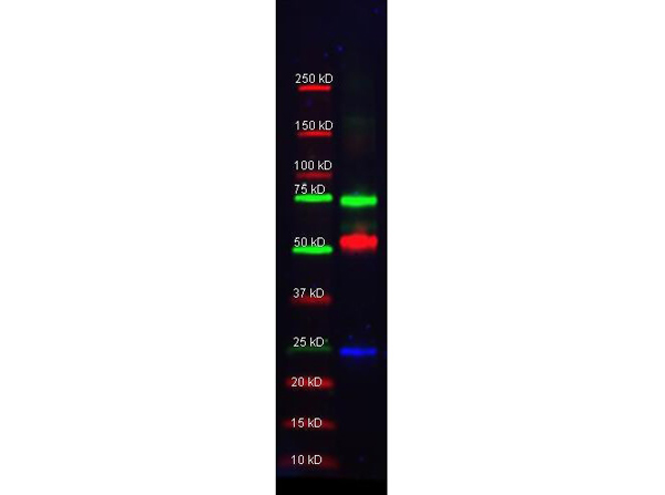
Multiplex western blot results using MB-070. Rockland Mouse-a-GST (200-301-200 lot 24882, blue), Rabbit anti-Transferrin (109-4134 lot 3033), and Goat-anti-Alpha-1-Anti-Trypsin (100-101-147 lot 5842) were used in a multiplex system to detect target proteins under reducing conditions in albumin depleted human serum with 320 ng of added GST. Sample was run by SDS-PAGE, transferred to 0.2 um PVDF using the BioRad Trans-Blot Turbo and blocked in 2.5% Blotto, 2.5% BSA, 0.02% Tween over night at 4°C. Membrane was probed with three primary antibodies at 1:1000 dilution (in MB-070 over night at 4°C). Detection shown was using DyLight?549 Donkey anti-Rabbit IgG (611-742-127 lot 21100, shown as green) DyLight?488 Donkey anti-Mouse IgG (610-741-124 lot 21095, shown as blue), and DyLight?649 Donkey anti-Goat IgG (605-743-125 lot 20834, shown as red) at 1:10,000 (in MB-070 at 30 min RT). Blots were washed, rinsed in methanol, dried and Images were collected using the BioRad VersaDoc System.
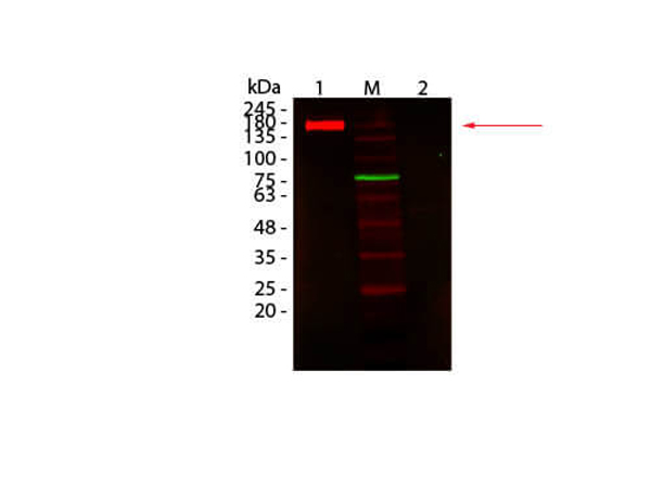
Western Blot of Fluorescent TrueBlotR: Anti-Rabbit IgG DyLight 680 Conjugated using MB-070. Lane 1: Rabbit IgG, Non-denatured. Lane 2: Rabbit IgG, Denatured. Load: 50 ng per lane. Primary antibody: none. Secondary antibody: Fluorescent TrueBlotR: Anti-Rabbit IgG DyLight 680 Conjugated antibody at 1:1,000 for 60 min at RT. Block: MB-070 for 30 min at RT. Predicted: 160 kDa for non-denatured; observed: 170-180 kDa for non-denatured. Band migrates at slightly higher molecular weight.
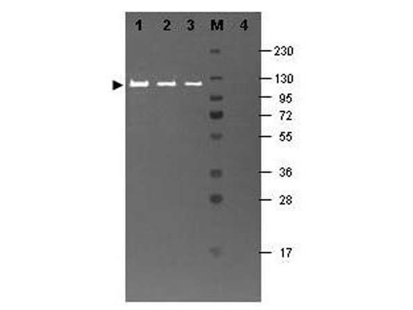
Western blot results using MB-070 and Fluorescein conjugated anti-b-Galactosidase antibody shows a band at ~117 kDa. Lanes 1 - 3 loaded with 60 ng, 30 ng and 15 ng, respectively of b-Gal present in partially purified preparations (arrowhead). Lane 4 shows no cross reactivity with proteins present in a non-specific control E.coli lysate. Proteins were resolved on a 4-20% Tris-Glycine gel by SDS-PAGE and transferred to nitrocellulose and blocking using Blocking Buffer for Fluorescent Western Blotting (p/n MB-070). The membrane was probed with fluorescein conjugated anti-b-Galactosidase (p/n 200-4236) diluted to 1:10,000. Reaction occurred for 2 hours at room temperature. Molecular weight estimation was made by comparison to a prestained MW marker in lane M. Fluorescence image was captured using the VersaDocR Imaging System developed by BIO-RAD. Other detection systems will yield similar results.
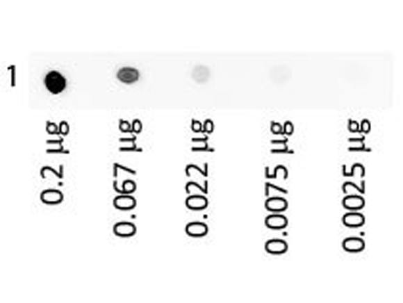
Dot Blot of Human IgA Fluorescein using MB-070. Antigen: Human IgA Fluorescein. Load: 3-fold serial dilution starting at 200 ng. Block: MB-070 for 30 min at RT.
702 Peptides are printed in duplicates randomly distributed on the microarray. Control peptides (HA and FLAG controls) are located in a square surrounding the peptides of interest. As secondary antibody DyLight? 549 conjugated goat anti-human IgG antibody and for the FLAG control peptide a mouse anti-FLAG-Cy3 antibody were used; microarrays were read using a Fujifilm Life Science FLA-5100 imaging system with a SHG 532nm (green) diode laser and an LPG filter. Fig e1. PMID:?26894206.
|
|
|
|
Blocking Buffer for Fluorescent Western Blotting
|
|
| 別品名 |
Multiplex Blocking Buffer, Fluorescent Blocking Buffer, Blocking Solution, Blocking Buffer Western Blot, IRDye Western Blot Blocking Buffer, Alexa Dye Blocking Buffer, DyLight Blocking Buffer
|
| 適用 |
Western Blot
|
| 標識物 |
Unlabeled
|
| 使用目的 |
このブロッキングバッファーは、IRDyeTM標識抗体を用いたウェスタンブロッティング用に特別にデザインされています。ご使用にあたってはニトロセルロースメンブレンを推奨しています。
|
| 参考文献 |
[Pub Med ID]33903110
|
|
| メーカー |
品番 |
包装 |
|
RKL
|
MB-070S
|
125 ML
|
※表示価格について
| 当社在庫 |
なし
|
| 納期目安 |
約10日
|
| 法規制 |
劇
|
| 保存温度 |
4℃禁凍結
|
|
※当社では商品情報の適切な管理に努めておりますが、表示される法規制情報は最新でない可能性があります。
また法規制情報の表示が無いものは、必ずしも法規制に非該当であることを示すものではありません。
商品のお届け前に最新の製品法規制情報をお求めの際はこちらへお問い合わせください。
|
※当社取り扱いの試薬・機器製品および受託サービス・創薬支援サービス(納品物、解析データ等)は、研究用としてのみ販売しております。
人や動物の医療用・臨床診断用・食品用としては、使用しないように、十分ご注意ください。
法規制欄に体外診断用医薬品と記載のものは除きます。
|
|
※リンク先での文献等のダウンロードに際しましては、掲載元の規約遵守をお願いします。
|
|
※CAS Registry Numbers have not been verified by CAS and may be inaccurate.
|
