|
※サムネイル画像をクリックすると拡大画像が表示されます。
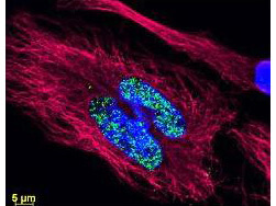
DyLightTM dyes can be used for multi color immunofluorescence microscopy with uniform fluorescence intensity throughout the image. DyLightTM dyes are exceptionally bright and photostable and are optimized for microscopy and microarray detection methods. This image shows anti histone detection using a DyLightTM 488 conjugate (green). Anti Tubulin was detected using a DyLightTM 549 conjugate (red). Nuclei were counter stained using DAPI (blue). The image was captured using an Axio Imager.Z1 (Zeiss Micro Imaging Inc).
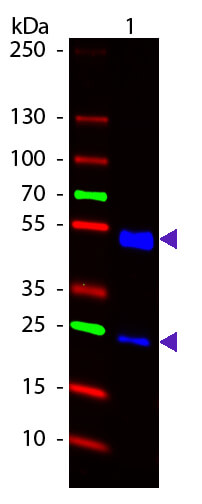
Western Blot of DylightTM 488 conjugated Goat Anti-Rat IgG secondary antibody. Lane 1: Rat IgG. Lane 2: None. Load: 50 ng per lane. Primary antibody: None. Secondary antibody: DylightTM 488 goat secondary antibody at 1:1,000 for 60 min at RT. Block: MB-070 for 30 min t RT. Predicted/Observed size: 25 & 55 kDa, 25 & 55 kDa for Rat IgG. Other band(s): None.
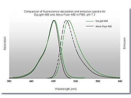
DyLightTM 488 Fluorescence Spectra
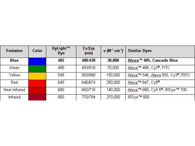
Properties of DyLightTM Fluorescent Dyes.
|


