|
※サムネイル画像をクリックすると拡大画像が表示されます。
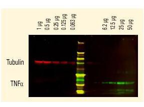
DyLightTM dyes can be used for two color Western Blot detection with low background and high signal. Anti tubulin was detected using a DyLightTM 680 conjugate. Anti TNFa was detected using a DyLightTM 800 conjugate. The image was captured using the OdysseyR Infrared Imaging System developed by LI COR.

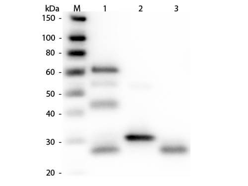
Western Blot of Unconjugated Anti-Chicken IgG (H&L) (RABBIT) Antibody (p/n 603-4102). Lane M: 3 μl Molecular Ladder. Lane 1: Chicken IgG whole molecule (p/n 003-0102). Lane 2: Chicken IgG F(c) Fragment (p/n 003-0103). Lane 3: Chicken IgG Fab Fragment (p/n 003-0105). All samples were reduced. Load: 50 ng per lane. Block: MB-070 for 30 min at RT. Primary Antibody: Anti-Chicken IgG (H&L) (RABBIT) Antibody (p/n 603-4102) 1:3,000 for 60 min at RT. Secondary antibody: Anti-Rabbit IgG (GOAT) Peroxidase Conjugated Antibody (p/n 611-103-122) 1:40,000 in MB-070 for 30 min at RT. Predicted/Observed Size: 25 and 72 kDa for Chicken IgY, 25 kDa for F(c) and Fab. Chicken F(c) migrates slightly higher.

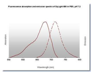
DyLightTM 680 Fluorescence Spectra.

Properties of DyLightTM Fluorescent Dyes.

|


