|
※サムネイル画像をクリックすると拡大画像が表示されます。
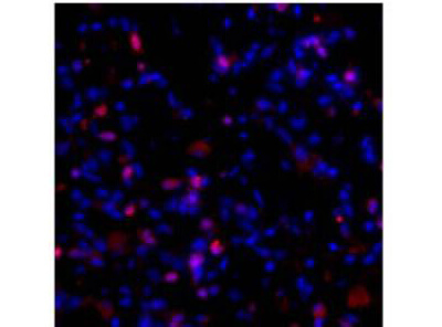
Immunofluorescence Microscopy of Rabbit Anti-Collagen I -alpha 1-propeptide antibody. Tissue: fibrotic mouse lung tissue. Fixation: 0.5% PFA. Antigen retrieval: not required. Primary antibody: Collagen I antibody at 10 μg/mL for 1 h at RT. Secondary antibody: Fluorescein rabbit secondary antibody at 1:10,000 for 45 min at RT. Localization: Collagen I is an extracellular matrix protein. Staining: Collagen I molecules (red) with bis-benzimide (blue) nuclear counterstain.
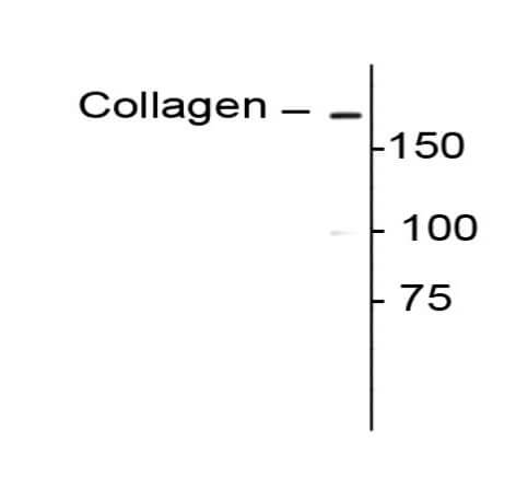
Western Blot of Rabbit anti-Collagen I alpha 1 propeptide antibody. Lane 1: rat lung lysate. Lane 2: none. Load: 10 μg per lane. Primary antibody: Collagen I antibody at 1:400 for overnight at 4°C. Secondary antibody: IRDye800TM rabbit secondary antibody at 1:10,000 for 45 min at RT. Block: 5% BLOTTO overnight at 4°C. Predicted/Observed size: ~180 kDa for collagen 1. Other band(s): none.
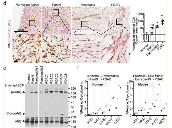
d)?Double-label immunohistochemical staining showing that anti-C-pro/α1(I) staining (red, with RL antibody) is in the extracellular space (negative for the cytoplasmic marker VIM in brown) of the mesenchymal regions in PDAC and pancreatitis samples but not in normal or PanIN pancreas. The mesenchymal regions are enlarged and quantified. The quantification was done by calculating the fraction of C-pro/α1(I) positive and VIM-negative area to that of VIM-negative ECM region and then normalizing the ratios to that of normal pancreas. Arrows point to extracellular C-pro/α1(I) regions. The numbers of quantified samples are 3, 3, 3, 7 (left to right). *p?<?0.05; **p?<?0.01; ns not significant. Two-tailed?t-tests are performed. All columns are represented by mean?±?SD.?e)?Western blotting (WB) showing large majority of the C-pro/α1(I) in the diseased pancreas ECM is present as uncleaved forms attached to α1(I), showing a representative image of three experimental repeats.?f)?Estimation of the percentage of each fibrillar collagen C-prodomain that is retained in the ECM in each stage. Note that there is increased C-prodomain retention as disease progresses. The percentages were derived from the average intensity of [1] peptides that map to the C-prodomain region divided by that of [2] peptides from the mature collagen region. Fig 1. PMID:?33879793
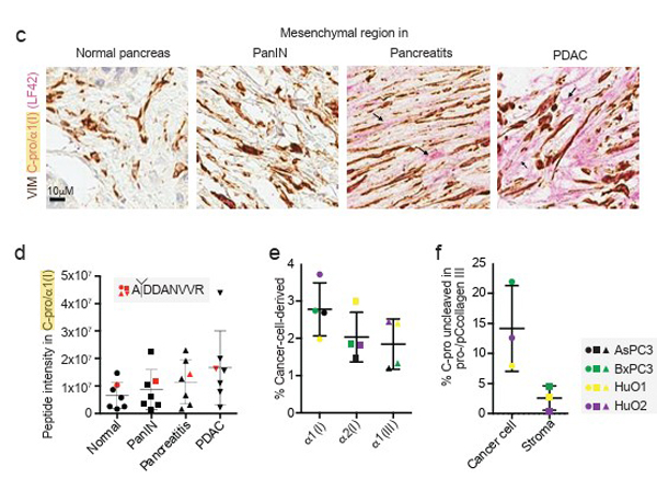
C), double color immunohistochemical staining showing that a-C-pro/a1(I) (LF42) staining is present in the extracellular space (negative for the cytoplasm marker VIM) in PDAC but not in normal pancreas in human. Arrows point to extracellular C-pro/a1(I) regions. The images are representative of 3, 3, 3, 7 tissue samples (left to right). D), intensity of peptides that map to human C-pro/a1(I) (N=7). Red marks represent the peptide that spans the pC-protease cleavage site (sequence shown) in human proa1(I). SF1. PMID:?33879793
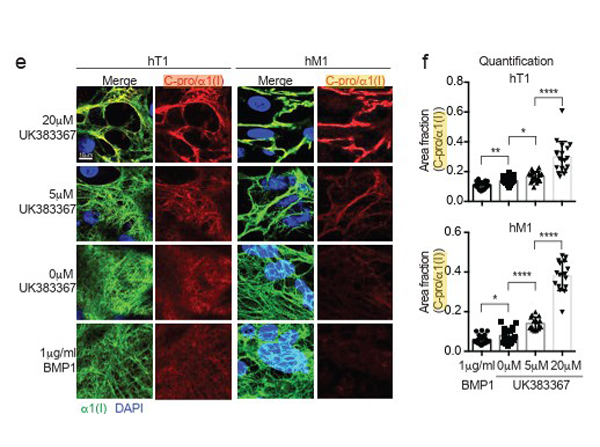
E, F) increasing amounts of BMP1 inhibitor UK383367 increased extracellular C-prodomain (Cpro/a1(I)) signal (E) after staining without permeabilization, quantified by the area fraction of mature a1(I) that is also positive for C-pro/a1(I) (F). On the contrary, adding BMP1 protein decreased C- pro/a1(I) signal (E,F). N numbers are 18, 17, 20, 19 for hT1 set and 20, 20, 15, 17 for hM1 set. SF2.PMID:?33879793
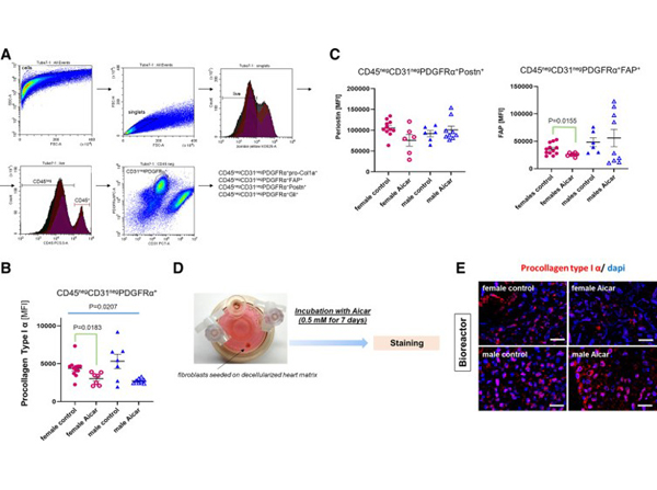
Collagen deposition and turnover in the old female and old male mouse heart in response to 5-aminoimidazole-4-carboxamide riboside (AICAR) treatment.?A: gating strategy to identify the nonmyocyte/nonmyeloid/nonendothelial-cell cardiac fibroblast population (CD45negCD31negPDGFRα+) that was analyzed for expression of procollagen type I α, fibroblast-activated protein (FAP), periostin (Postn), and Gli1.?B: mean fluorescence intensity (MFI) of procollagen type I α in CD45negCD31negPDGFRα+?cells isolated from control or AICAR-treated old female and male mouse hearts.?C: graph depicting the MFI for periostin (left) and FAP (right) in the CD45negCD31negPDGFRα+?cells (n?= 6?12/group).?D: summary of the ex vivo experiments performed using bioreactor vessels. Fibroblasts were seeded on sex-matched matrices isolated from old mouse hearts. They were then incubated with 0.5 mM AICAR for 7 days.?E: immunostaining for procollagen type 1α in cardiac fibroblasts seeded on sex-matched matrices in the bioreactor. Scale bar = 50 μm (n?= 3). Brown?Forsythe and Welch one-way ANOVA was used to test the significant difference among the four groups, to determine the significant difference among the groups for?B?and?C?(n?= 7?12). Fig 4. PMID:?35714177
|
|
|
|
Immunofluorescence Microscopy of Rabbit Anti-Collagen I -alpha 1-propeptide antibody. Tissue: fibrotic mouse lung tissue. Fixation: 0.5% PFA. Antigen retrieval: not required. Primary antibody: Collagen I antibody at 10 μg/mL for 1 h at RT. Secondary antibody: Fluorescein rabbit secondary antibody at 1:10,000 for 45 min at RT. Localization: Collagen I is an extracellular matrix protein. Staining: Collagen I molecules (red) with bis-benzimide (blue) nuclear counterstain.
|
|
| 別品名 |
Alpha-1 type I collagen, COLA1A1
|
| 交差種 |
Human
Mouse
Rat
|
| 適用 |
Western Blot
Immunohistochemistry
|
| 免疫動物 |
Rabbit
|
| 標識物 |
Unlabeled
|
| 精製度 |
Affinity Purified
|
| GENE ID |
1277
|
| Accession No.(Gene/Protein) |
NP_000079.2, P02452.5
|
| Gene Symbol |
COL1A1
|
| 参考文献 |
[[Cosmo Reference Collection]][Pub Med ID]32277974,33879793,32075905,35714177
|
|
| メーカー |
品番 |
包装 |
|
RKL
|
600-401-D19
|
100 UL
|
※表示価格について
| 当社在庫 |
なし
|
| 納期目安 |
約10日
|
| 保存温度 |
-20℃
|
|
※当社では商品情報の適切な管理に努めておりますが、表示される法規制情報は最新でない可能性があります。
また法規制情報の表示が無いものは、必ずしも法規制に非該当であることを示すものではありません。
商品のお届け前に最新の製品法規制情報をお求めの際はこちらへお問い合わせください。
|
※当社取り扱いの試薬・機器製品および受託サービス・創薬支援サービス(納品物、解析データ等)は、研究用としてのみ販売しております。
人や動物の医療用・臨床診断用・食品用としては、使用しないように、十分ご注意ください。
法規制欄に体外診断用医薬品と記載のものは除きます。
|
|
※リンク先での文献等のダウンロードに際しましては、掲載元の規約遵守をお願いします。
|
|
※CAS Registry Numbers have not been verified by CAS and may be inaccurate.
|


