|
※サムネイル画像をクリックすると拡大画像が表示されます。
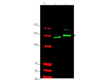
Western blot using Rockland's affinity purified anti-Gli-2 antibody shows detection of Gli-2 protein. Lane 1: rat testes (p/n W12-000-GZ3) and Lane 2: human HEK293 (p/n W09-000-365) whole cell lysates (arrowhead).? See Ruppert et al for testing conditions.? Each lane contains approximately 35μg of lysate.? Primary antibody was used at a 1:400 dilution in 5% BLOTTO (p/n B501-0500) in PBS overnight at 4°C.? The membrane was washed and reacted with a 1:10,000 dilution of IRDyeR 800 conjugated Gt-a-Rabbit IgG [H&L] MX10 (p/n 611-132-122) for 45 min at room temperature (800 nm channel, green).?? Molecular weight estimation was made by comparison to prestained MW markers in lane M (700 nm channel, red).?? IRDyeR 800 fluorescence image was captured using the OdysseyR Infrared Imaging System developed by LI-COR. IRDye is a trademark of LI-COR, Inc.? Other detection systems will yield similar results.
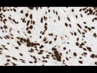
Rockland's Affinity Purified anti-Gli2 antibody shows strong cytoplasmic and membranous staining of tumor cells in human breast tissue. Tissue was formalin-fixed and paraffin embedded. Brown color indicates presence of protein, blue color shows cell nuclei. Personal Communication, Kenneth Wester, www.proteinatlas.org, Uppsala, Sweden.
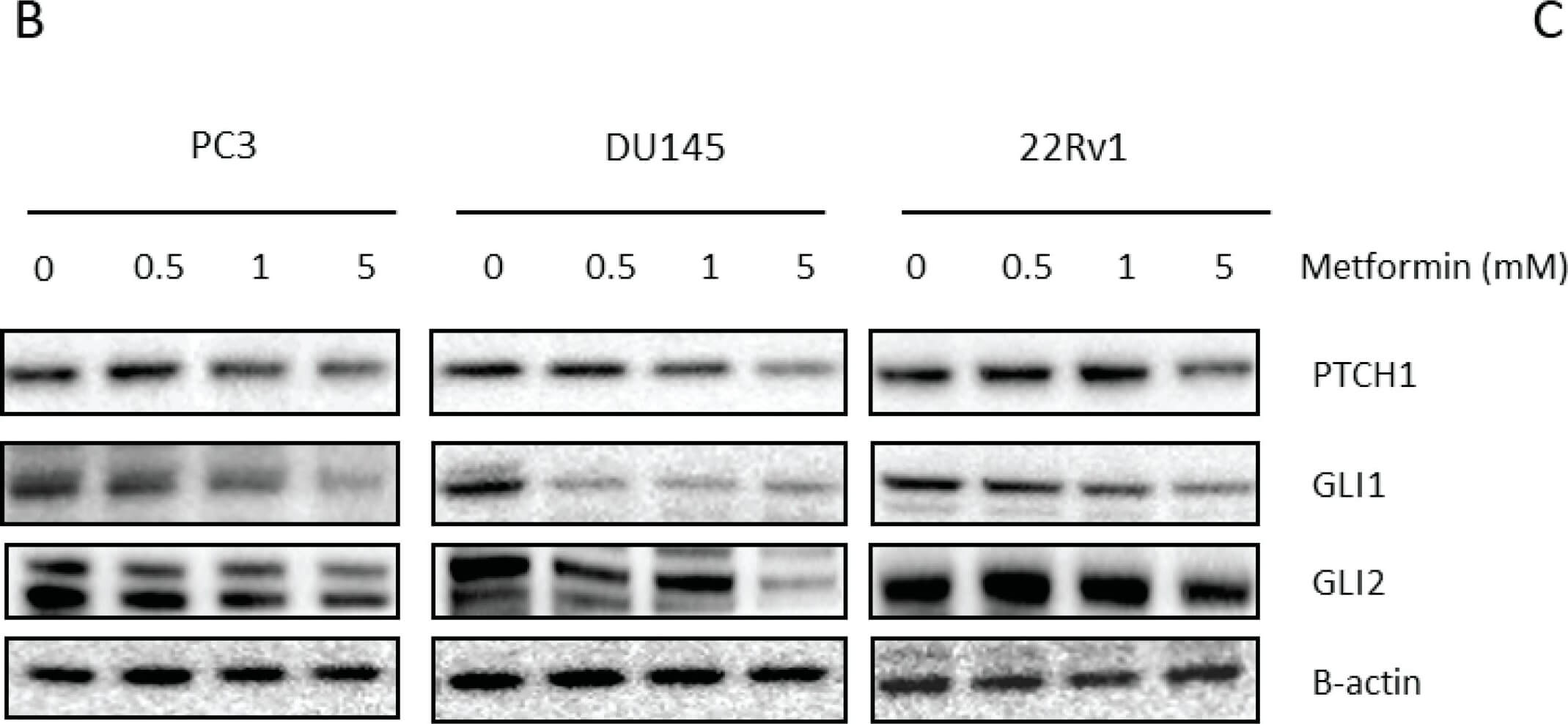
Link between metformin and Hedgehog signaling. (A) GLI1, GLI2 and PTCH1 gene expression after 72-h metformin treatment. Means ± SEM of two independent experiments. * p < 0.05 vs. control; (B) PTCH1, GLI1 and GLI2 protein expression after 72-h metformin treatment; (C) (p)AMPK protein and GLI1 expression in 22Rv1 cells transfected with AMPK siRNA and treated with metformin (5 mM) 72-h prior to protein lysis. GLI1, glioma-associated oncogene homolog 1; GLI2, glioma-associated oncogene homolog 2; PTCH1, patched 1. Figure provided by CiteAb. Source: Int J Mol Sci, PMID: 28208838.
|
|
|
|
Western blot using Rockland's affinity purified anti-Gli-2 antibody shows detection of Gli-2 protein. Lane 1: rat testes (p/n W12-000-GZ3) and Lane 2: human HEK293 (p/n W09-000-365) whole cell lysates (arrowhead).? See Ruppert et al for testing conditions.? Each lane contains approximately 35μg of lysate.? Primary antibody was used at a 1:400 dilution in 5% BLOTTO (p/n B501-0500) in PBS overnight at 4°C.? The membrane was washed and reacted with a 1:10,000 dilution of IRDyeR 800 conjugated Gt-a-Rabbit IgG [H&L] MX10 (p/n 611-132-122) for 45 min at room temperature (800 nm channel, green).?? Molecular weight estimation was made by comparison to prestained MW markers in lane M (700 nm channel, red).?? IRDyeR 800 fluorescence image was captured using the OdysseyR Infrared Imaging System developed by LI-COR. IRDye is a trademark of LI-COR, Inc.? Other detection systems will yield similar results.
|
|
| 別品名 |
rabbit anti-GLI-2 antibody, GLI2, zinc finger protein GLI2, GLI family zinc finger protein 2, Tax helper protein antibody, THP antibody
|
| 交差種 |
Human
Rat
|
| 適用 |
Western Blot
Enzyme Linked Immunosorbent Assay
Immunohistochemistry
|
| 免疫動物 |
Rabbit
|
| 抗原部位 |
a.a.46-60
|
| 標識物 |
Unlabeled
|
| 精製度 |
Affinity Purified
|
| GENE ID |
2736
|
| Accession No.(Gene/Protein) |
NP_005261.2, P10070
|
| Gene Symbol |
GLI2
|
| 参考文献 |
[Pub Med ID]27713179
|
| [注意事項] |
濃度はロットによって異なる可能性があります。メーカーDS及びCoAからご確認ください。
|
|
| メーカー |
品番 |
包装 |
|
RKL
|
600-401-845S
|
25 UL
|
※表示価格について
| 当社在庫 |
なし
|
| 納期目安 |
約10日
|
| 保存温度 |
-20℃
|
|
※当社では商品情報の適切な管理に努めておりますが、表示される法規制情報は最新でない可能性があります。
また法規制情報の表示が無いものは、必ずしも法規制に非該当であることを示すものではありません。
商品のお届け前に最新の製品法規制情報をお求めの際はこちらへお問い合わせください。
|
※当社取り扱いの試薬・機器製品および受託サービス・創薬支援サービス(納品物、解析データ等)は、研究用としてのみ販売しております。
人や動物の医療用・臨床診断用・食品用としては、使用しないように、十分ご注意ください。
法規制欄に体外診断用医薬品と記載のものは除きます。
|
|
※リンク先での文献等のダウンロードに際しましては、掲載元の規約遵守をお願いします。
|
|
※CAS Registry Numbers have not been verified by CAS and may be inaccurate.
|
