

| 別品名 |
Arresten antibody, Canstatin antibody, Collagen Of Basement Membrane Alpha 1 Chain antibody
|
| 種由来 |
Mammalian
|
| 由来詳細 |
[Species]Human/Bovine
|
| 標識物 |
Unlabeled
|
| 精製度 |
Affinity Purified
|
| 適用 |
Immunohistochemistry
Dot Blot
|
| 免疫動物 |
Rabbit
|
| 抗体クラス |
IgG
|
| 交差種 |
Human
Bovine
|
| GENE ID |
1282
|
| Accession No.(Gene/Protein) |
NP_001290039, P02462
|
| Gene Symbol |
COL4A4
|
| 形状 |
滅菌済み液状品
|
| 参考文献 |
[Pub Med ID]33239448, 32409724, 27444354, 27687499, 28877431, 29769449, 23922030, 32478392, 21330653, 21330653, 24529431, 29540474, 30388216, 27815317, 24284212, 24713487, 11493641, 21697382, 22485136, 22925884+他多数
|
| [注意事項] |
濃度はロットによって異なる可能性があります。メーカーDS及びCoAからご確認ください。
|
|
※サムネイル画像をクリックすると拡大画像が表示されます。
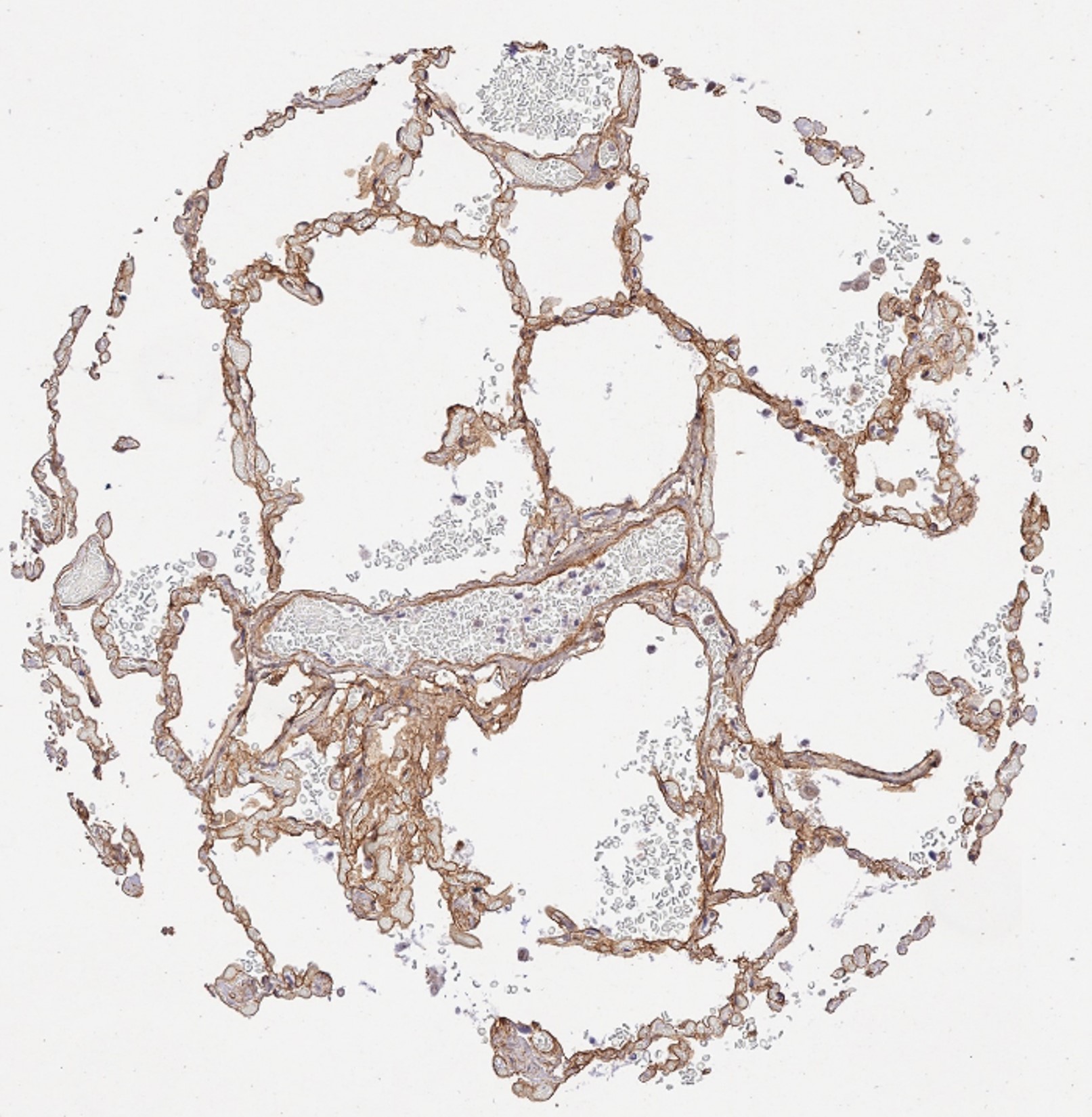
Immunohistochemistry results of Rabbit Anti Collagen Type IV Antibody. Tissue: human lung tissue. Fixation: FFPE. Antigen Retrieval: HIER using Tris EDTA citrate buffer pH 7.8 for 5 min. Blocking: Peroxidase Blocking Solution for 10 min. Primary Antibody: Anti Collagen Type IV (p/n 600 401 106 0.1) at 1:15 for 1 hr at 37 C. Secondary Antibody: Dako REAL EnVision Detection Kit, Polymer HRP, Rabbit/Mouse. Counterstain: Hematoxylin for 15 sec.Substrate: DAB Chromogen, Rabbit/Mouse. Staining/Results: basement membranes and vessels.
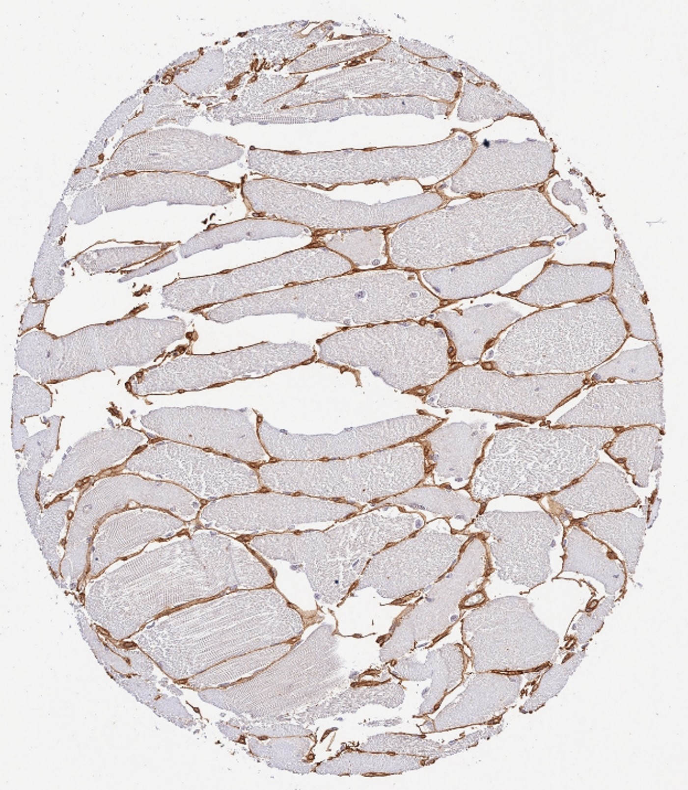
Immunohistochemistry results of Rabbit Anti-Collagen Type IV Antibody. Tissue: human skeletal muscle cells. Fixation: FFPE. Antigen Retrieval: HIER using Tris-EDTA-citrate buffer pH 7.8 for 5 min. Blocking: Peroxidase-Blocking Solution for 10 min. Primary Antibody: Anti-Collagen Type IV (p/n 600-401-106-0.1) at 1:15 for 1 hr at 37 C. Secondary Antibody: Dako REAL EnVision Detection Kit, Polymer-HRP, Rabbit/Mouse. Counterstain: Hematoxylin for 15 sec.Substrate: DAB-Chromogen, Rabbit/Mouse. Staining/Results: cells surrounded by collagen IV fibers.
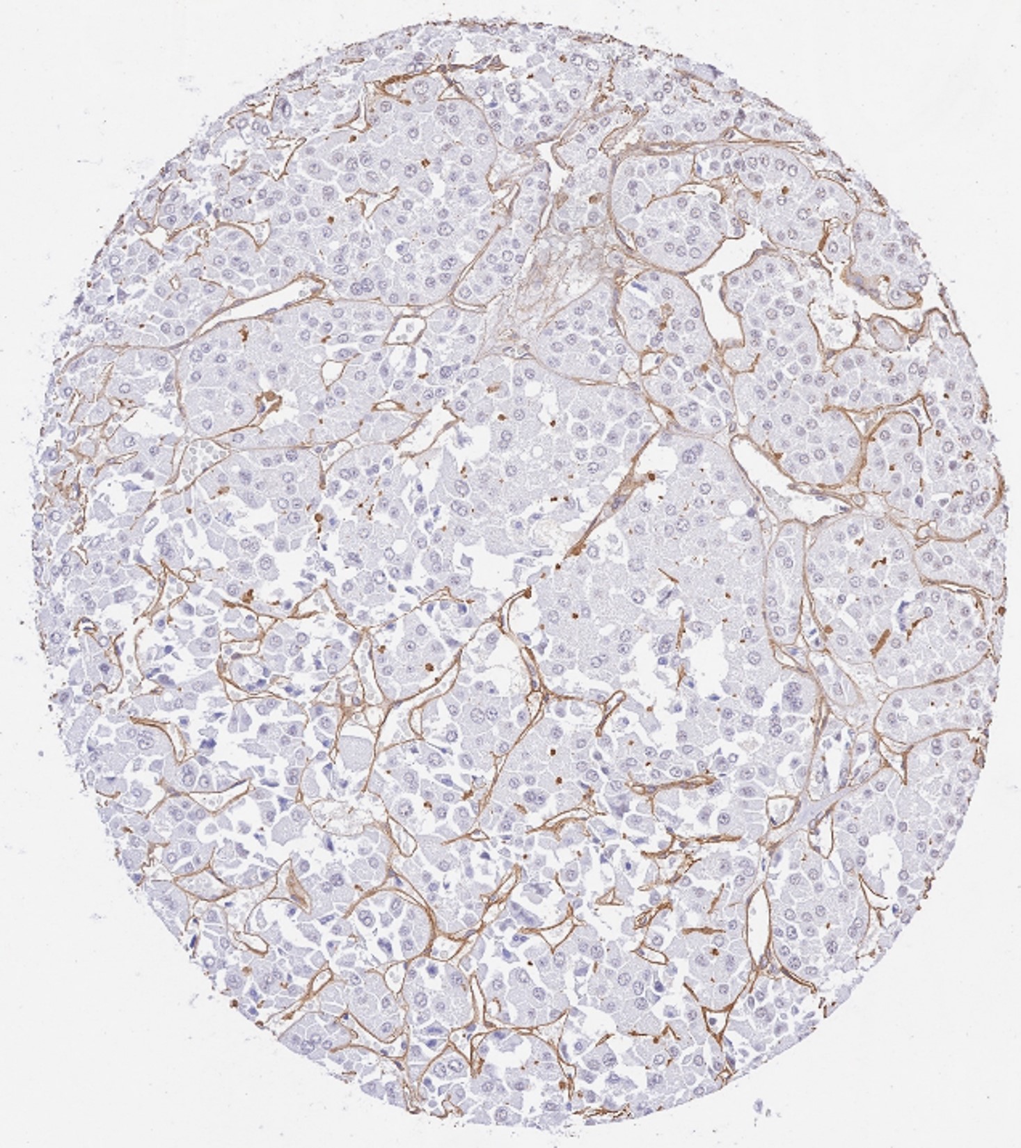
Immunohistochemistry results of Rabbit Anti-Collagen Type IV Antibody. Tissue: human renal oncocytoma. Fixation: FFPE. Antigen Retrieval: HIER using Tris-EDTA-citrate buffer pH 7.8 for 5 min. Blocking: Peroxidase-Blocking Solution for 10 min. Primary Antibody: Anti-Collagen Type IV (p/n 600-401-106-0.1) at 1:15 for 1 hr at 37 C. Secondary Antibody: Dako REAL EnVision Detection Kit, Polymer-HRP, Rabbit/Mouse. Counterstain: Hematoxylin for 15 sec.Substrate: DAB-Chromogen, Rabbit/Mouse. Staining/Results: dense collagen IV positive membranes surrounding tumor cell nests.
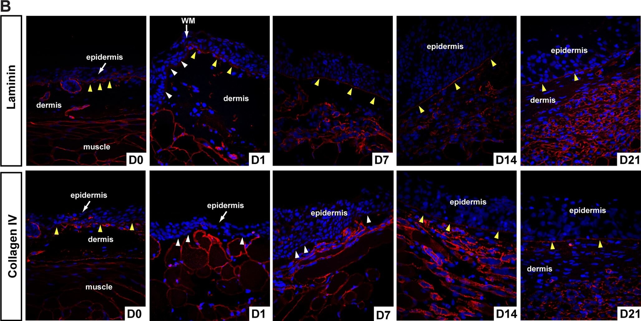
Lamina lucida and lamina densa regenerate before new ECM deposition.A) Histological examination of basement membrane (BM) regeneration in axolotls. The uninjured BM is visible as a thick blue-stained fibrous band (yellow arrows). An immature BM has begun to reform (yellow arrow D1) after re-epithelialization and is visible at the wound margin (WM) in contrast to the uninjured BM. The regenerated BM is visible at D47. Yellow arrows at D7 and D21 indicate reforming BM. B) Examination of lamina lucida (laminin) and lamina densa (collagen type IV) during basement membrane regeneration. The uninjured BM is positive for laminin and collagen type IV (yellow arrows) as are the basement membranes surrounding glands and muscle fibers. Following re-epithelialization the basal lamina of the epidermis is negative for laminin and collagen type IV (white arrows) and this is clearly evident at the wound margin (WM). Seven days post injury the BM stains strongly for laminin indicating reformation of the lamina lucida, while staining for collagen type IV is punctuated. The lamina densa is regenerated by D14 based on continuous collagen type IV staining and persists during dermal regeneration. Figure provided by CiteAb. Source: PLoS One, PMID: 22485136.
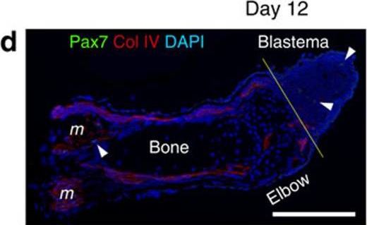
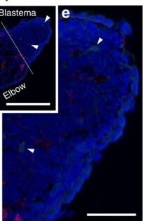
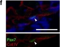
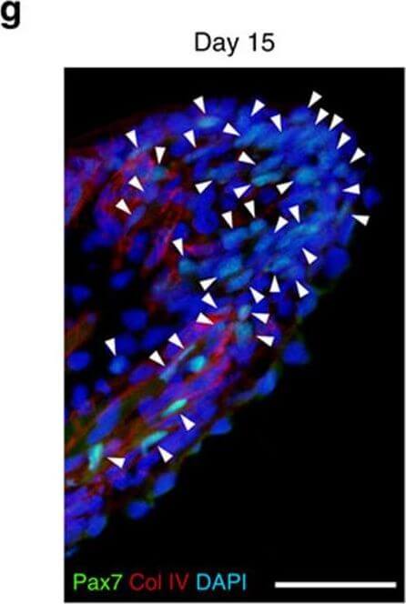
SMFC tracking in larval newt limb regeneration.(a) Larva (3 months old). It has four limbs, as well as the gills and tail fin. Scale bar, 4?mm. (b) Monitoring of SMFCs (mCherry+) during limb regeneration (n=6). mCherry was not detected in the regenerating part of the limb until ?30 days when the amputated limb had almost been recovered (see Supplementary Movie 1). Arrowheads: flexor muscle for digits (see Fig. 2). Scale bar, 1?mm. (c) Sections of regenerating limbs (n=3 for each stage). SMFC-derived mCherry+ cells were not observed in the blastema. Lines: amputation site. m: muscle. Scale bar, 100?μm. (d?f) Pax7 immunolabelling of regenerating limbs on day 12 (n=3) and (g) day 15 (n=3) after amputation. (d) On day 12, a few Pax7+ nuclei (arrowheads) were detected in blastema cells and in satellite cells along the muscle fibres. Col IV, collagen type IV immunoreactivity. DAPI (4,6-diamidino-2-phenylindole), nuclei. Scale bar, 300?μm. The Pax7+ nuclei pointed by arrowheads were enlarged in e and f, respectively. Scale bars, 100?μm. (g) On day 15 when the regenerating part of the limb grew more distally, the number of Pax7+ nuclei (arrowheads) in the blastema was dramatically increased. Scale bar, 100?μm. (h) Summary. In larval newts, MPCs, potentially satellite cells, were recruited for new muscle during limb regeneration, whereas SMFCs were not. Figure provided by CiteAb. Source: Nat Commun, PMID: 27026263.
SMFC tracking in larval newt limb regeneration.(a) Larva (3 months old). It has four limbs, as well as the gills and tail fin. Scale bar, 4mm. (b) Monitoring of SMFCs (mCherry+) during limb regeneration (n=6). mCherry was not detected in the regenerating part of the limb until 30 days when the amputated limb had almost been recovered (see Supplementary Movie 1). Arrowheads: flexor muscle for digits (see Fig. 2). Scale bar, 1?mm. (c) Sections of regenerating limbs (n=3 for each stage). SMFC-derived mCherry+ cells were not observed in the blastema. Lines: amputation site. m: muscle. Scale bar, 100?μm. (d?f) Pax7 immunolabelling of regenerating limbs on day 12 (n=3) and (g) day 15 (n=3) after amputation. (d) On day 12, a few Pax7+ nuclei (arrowheads) were detected in blastema cells and in satellite cells along the muscle fibres. Col IV, collagen type IV immunoreactivity. DAPI (4,6-diamidino-2-phenylindole), nuclei. Scale bar, 300?μm. The Pax7+ nuclei pointed by arrowheads were enlarged in e and f, respectively. Scale bars, 100?μm. (g) On day 15 when the regenerating part of the limb grew more distally, the number of Pax7+ nuclei (arrowheads) in the blastema was dramatically increased. Scale bar, 100?μm. (h) Summary. In larval newts, MPCs, potentially satellite cells, were recruited for new muscle during limb regeneration, whereas SMFCs were not. Figure provided by CiteAb. Source: Nat Commun, PMID: 27026263.
SMFC tracking in larval newt limb regeneration.(a) Larva (3 months old). It has four limbs, as well as the gills and tail fin. Scale bar, 4?mm. (b) Monitoring of SMFCs (mCherry+) during limb regeneration (n=6). mCherry was not detected in the regenerating part of the limb until 30 days when the amputated limb had almost been recovered (see Supplementary Movie 1). Arrowheads: flexor muscle for digits (see Fig. 2). Scale bar, 1?mm. (c) Sections of regenerating limbs (n=3 for each stage). SMFC-derived mCherry+ cells were not observed in the blastema. Lines: amputation site. m: muscle. Scale bar, 100?μm. (d?f) Pax7 immunolabelling of regenerating limbs on day 12 (n=3) and (g) day 15 (n=3) after amputation. (d) On day 12, a few Pax7+ nuclei (arrowheads) were detected in blastema cells and in satellite cells along the muscle fibres. Col IV, collagen type IV immunoreactivity. DAPI (4,6-diamidino-2-phenylindole), nuclei. Scale bar, 300?μm. The Pax7+ nuclei pointed by arrowheads were enlarged in e and f, respectively. Scale bars, 100?μm. (g) On day 15 when the regenerating part of the limb grew more distally, the number of Pax7+ nuclei (arrowheads) in the blastema was dramatically increased. Scale bar, 100?μm. (h) Summary. In larval newts, MPCs, potentially satellite cells, were recruited for new muscle during limb regeneration, whereas SMFCs were not. Figure provided by CiteAb. Source: Nat Commun, PMID: 27026263.
SMFC tracking in larval newt limb regeneration.(a) Larva (3 months old). It has four limbs, as well as the gills and tail fin. Scale bar, 4?mm. (b) Monitoring of SMFCs (mCherry+) during limb regeneration (n=6). mCherry was not detected in the regenerating part of the limb until 30 days when the amputated limb had almost been recovered (see Supplementary Movie 1). Arrowheads: flexor muscle for digits (see Fig. 2). Scale bar, 1?mm. (c) Sections of regenerating limbs (n=3 for each stage). SMFC-derived mCherry+ cells were not observed in the blastema. Lines: amputation site. m: muscle. Scale bar, 100?μm. (d?f) Pax7 immunolabelling of regenerating limbs on day 12 (n=3) and (g) day 15 (n=3) after amputation. (d) On day 12, a few Pax7+ nuclei (arrowheads) were detected in blastema cells and in satellite cells along the muscle fibres. Col IV, collagen type IV immunoreactivity. DAPI (4,6-diamidino-2-phenylindole), nuclei. Scale bar, 300?μm. The Pax7+ nuclei pointed by arrowheads were enlarged in e and f, respectively. Scale bars, 100?μm. (g) On day 15 when the regenerating part of the limb grew more distally, the number of Pax7+ nuclei (arrowheads) in the blastema was dramatically increased. Scale bar, 100?μm. (h) Summary. In larval newts, MPCs, potentially satellite cells, were recruited for new muscle during limb regeneration, whereas SMFCs were not. Figure provided by CiteAb. Source: Nat Commun, PMID: 27026263.
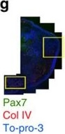
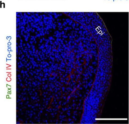
SMFC tracking in metamorphosed newt limb regeneration.(a) Juvenile (16 months old). Scale bar, 15?mm. (b) Limb regeneration. Scale bar, 5?mm. (c?e) Tracking of SMFCs (mCherry+) (n=2). This animal was a mosaic expressing EGFP in muscle only. mCherry+ fibres in the forearm were ?25% of total EGFP+ fibres. (c) On day 36 after amputation, fragments of muscle fibres (arrows) were observed in distal regions adjacent to the blastema. Scale bar, 100?μm. (d) mCherry+ mononucleated cells (red arrowheads; enlarged in right-hand panels) and EGFP+ cells (green arrowheads) were observed in the blastema. Epi, epidermis. To-pro-3: nuclei. Scale bars, 50?μm (left), 10?μm (right-hand panels). (e) In the same limb, at day 96 after the second amputation in the upper arm (line), mCherry (arrows) and EGFP were observed only in muscle fibres. Scale bars, 1?mm (upper panel), 500?μm (lower panels). (f?i) Pax7 immunolabelling of a regenerating limb on day 26 after amputation (n=4). Pax7 immunoreactivity was not detected in the blastema. (f) Translucent image. Line: amputation site. (g) Merged fluorescence image. Col IV, collagen type IV immunoreactivity. To-pro-3: nuclei. Scale bar, 1?mm. (h) Enlargement of a region in the blastema and (i) a region proximal to the amputation site, enclosed by boxes in g. Scale bars, 250?μm. Arrowheads in (i) Pax7+ nuclei. An example satellite cell (box) is enlarged in the right-hand panels (upper: Col VI/To-pro-3; lower: Col IV/Pax7). Scale bar, 50?μm. (j) Summary. In metamorphosed newts, SMFCs were recruited for new muscle during limb regeneration, whereas MPCs such as satellite cells were not. Figure provided by CiteAb. Source: Nat Commun, PMID: 27026263.
SMFC tracking in metamorphosed newt limb regeneration.(a) Juvenile (16 months old). Scale bar, 15?mm. (b) Limb regeneration. Scale bar, 5?mm. (c?e) Tracking of SMFCs (mCherry+) (n=2). This animal was a mosaic expressing EGFP in muscle only. mCherry+ fibres in the forearm were ?25% of total EGFP+ fibres. (c) On day 36 after amputation, fragments of muscle fibres (arrows) were observed in distal regions adjacent to the blastema. Scale bar, 100?μm. (d) mCherry+ mononucleated cells (red arrowheads; enlarged in right-hand panels) and EGFP+ cells (green arrowheads) were observed in the blastema. Epi, epidermis. To-pro-3: nuclei. Scale bars, 50?μm (left), 10?μm (right-hand panels). (e) In the same limb, at day 96 after the second amputation in the upper arm (line), mCherry (arrows) and EGFP were observed only in muscle fibres. Scale bars, 1?mm (upper panel), 500?μm (lower panels). (f?i) Pax7 immunolabelling of a regenerating limb on day 26 after amputation (n=4). Pax7 immunoreactivity was not detected in the blastema. (f) Translucent image. Line: amputation site. (g) Merged fluorescence image. Col IV, collagen type IV immunoreactivity. To-pro-3: nuclei. Scale bar, 1?mm. (h) Enlargement of a region in the blastema and (i) a region proximal to the amputation site, enclosed by boxes in g. Scale bars, 250?μm. Arrowheads in (i) Pax7+ nuclei. An example satellite cell (box) is enlarged in the right-hand panels (upper: Col VI/To-pro-3; lower: Col IV/Pax7). Scale bar, 50?μm. (j) Summary. In metamorphosed newts, SMFCs were recruited for new muscle during limb regeneration, whereas MPCs such as satellite cells were not. Figure provided by CiteAb. Source: Nat Commun, PMID: 27026263.
|
|
|
|
Immunohistochemistry results of Rabbit Anti Collagen Type IV Antibody. Tissue: human lung tissue. Fixation: FFPE. Antigen Retrieval: HIER using Tris EDTA citrate buffer pH 7.8 for 5 min. Blocking: Peroxidase Blocking Solution for 10 min. Primary Antibody: Anti Collagen Type IV (p/n 600 401 106 0.1) at 1:15 for 1 hr at 37 C. Secondary Antibody: Dako REAL EnVision Detection Kit, Polymer HRP, Rabbit/Mouse. Counterstain: Hematoxylin for 15 sec.Substrate: DAB Chromogen, Rabbit/Mouse. Staining/Results: basement membranes and vessels.
|
|
|
| メーカー |
品番 |
包装 |
|
RKL
|
600-401-106S
|
25 UL
|
※表示価格について
| 当社在庫 |
なし
|
| 納期目安 |
約10日
|
| 保存温度 |
-20℃
|
|

