|
※サムネイル画像をクリックすると拡大画像が表示されます。
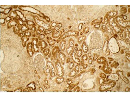
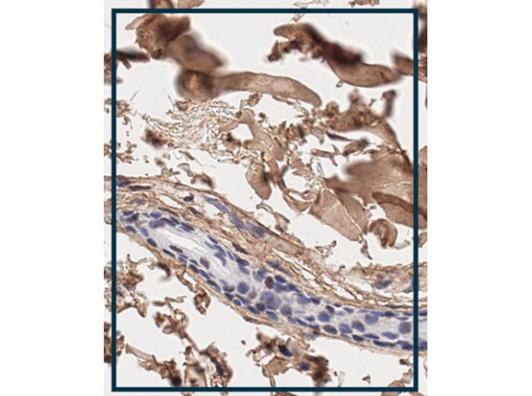
Rockland's Affinity Purified anti-Collagen I antibody was used at a 1:100 dilution to detect distal tubules in normal kidney tissue. Note the absence of staining of glomeruli. The antibody was reacted with antibody for 4 hours at room temperature followed by the addition of secondary antibody and substrate reaction. Tissue was formalin-fixed and paraffin embedded. No antigen retrieval was performed.
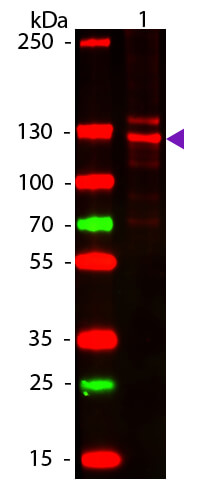
Western blot of Human Collagen Type I. Lane 1: Human Collagen Type 1 (p/n 009-001-103). Load: 50 ng per lane. Primary antibody: Collagen Type I antibody at 1:1,000 overnight at 4°C. Secondary antibody: DyLight? 649 rabbit secondary antibody (p/n 611-743-127) at 1:20,000 for 30 min at RT. Block: (p/n MB-070( for 30 min at RT. Predicted/Observed size: 139 & 130 kDa, 139 & 130 kDa for Collagen Type I. Other Band(s): Collagen Type I splice variants and isoforms.
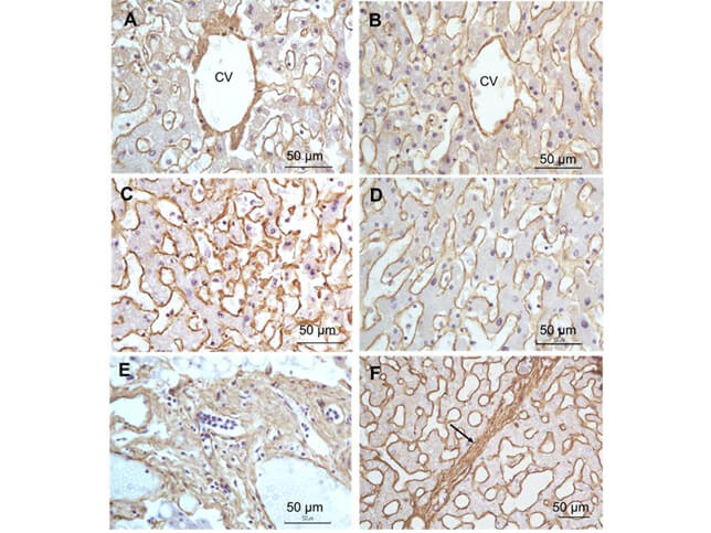
Immunohistochemistry of Rabbit Anti-collagen type I antibody. Tissue: right lobe of the liver section. A:Central Vein (CV) fibrosis, B: Non-fibrotic CV, C: Perisinusodial fibrosis, D: Non-fibrotic area, E: Protat tract fibrosis, F: Septal fibrosis (arrow). Fixation: formalin fixed paraffin embedded. Antigen retrieval: not required. Primary antibody: Anti-collagen type I at 1:1250 for 4°C for 24hr. Secondary antibody: Peroxidase biotin-streptavidin rabbit secondary antibody at 1:10,000 for 45 min at RT. Localization: Anti-collagen type I is intra and extracellular. Staining: 3.3’-diaminobenzidine tetrahydrochloride was used as the chromogen. Nuclei were counterstained purple with hematoxylin.
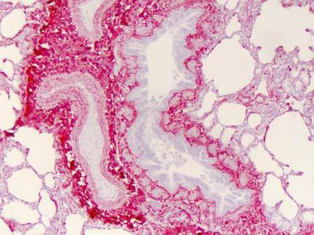
Immunohistochemistry of Collagen I antibody. Tissue: human lung. Fixation: formalin fixed paraffin embedded. Antigen retrieval: user optimized. Primary antibody: Collagen 1 1:400 Secondary antibody: Peroxidase goat anti-rabbit at 1:10,000 for 45 min at RT. Localization: Strong staining was observed in the extracellular matrix of the lung. Epithelial cells were negative. Staining: antibody as precipitated red signal with a hematoxylin purple nuclear counterstain.
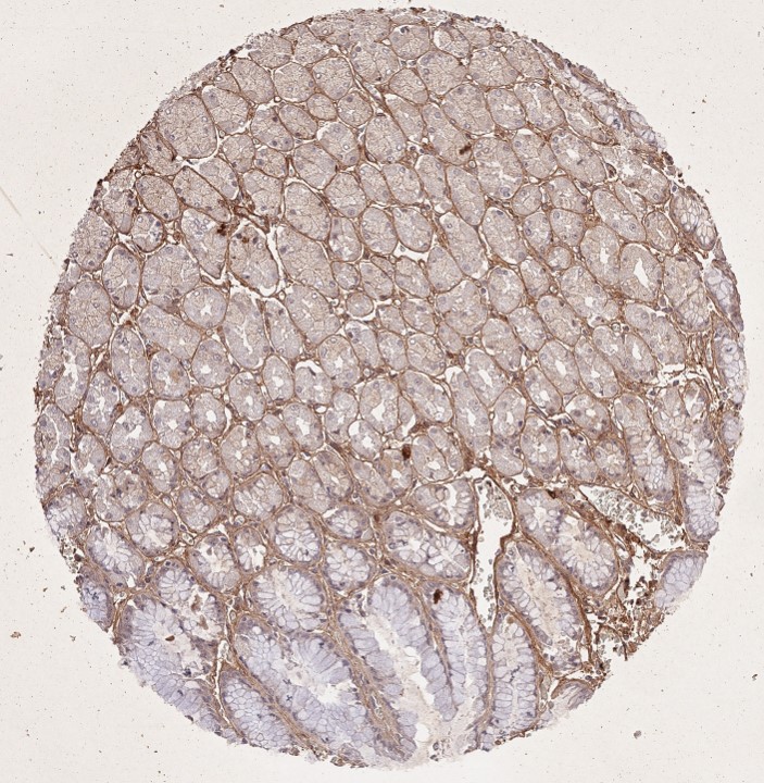
Immunohistochemistry results of Rabbit Anti-Collagen Type I Antibody. Tissue: human stomach mucosa (TMA). Fixation: FFPE. Antigen Retrieval: HIER using Tris-EDTA-citrate buffer pH 7.8 for 5 min. Blocking: Peroxidase-Blocking Solution for 10 min. Primary Antibody: Anti-Collagen Type I (p/n 600-401-103-0.1) at 1:15 for 1 hr at 37 °C. Secondary Antibody: Dako REAL EnVision Detection Kit, Polymer-HRP, Rabbit/Mouse. Counterstain: Hematoxylin for 15 sec.Substrate: DAB-Chromogen, Rabbit/Mouse. Staining/Results: basement membranes and blood vessels. Independently Validated by?antibodies-online GmbH (p/n ABIN7565871/ ABIN5596819/ ABIN5596820) by?MS Validated Antibodies.
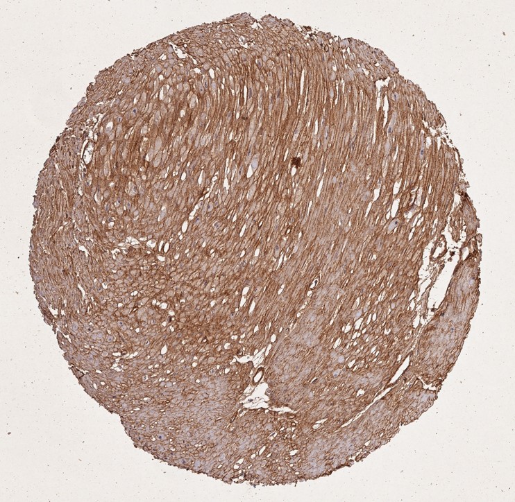
Immunohistochemistry results of Rabbit Anti-Collagen Type I Antibody. Tissue: smooth muscle cells of human stomach wall. Fixation: FFPE. Antigen Retrieval: HIER using Tris-EDTA-citrate buffer pH 7.8 for 5 min. Blocking: Peroxidase-Blocking Solution for 10 min. Primary Antibody: Anti-Collagen Type I (p/n 600-401-103-0.1) at 1:15 for 1 hr at 37 °C. Secondary Antibody: Dako REAL EnVision Detection Kit, Polymer-HRP, Rabbit/Mouse. Counterstain: Hematoxylin for 15 sec.Substrate: DAB-Chromogen, Rabbit/Mouse. Staining/Results: smooth muscle cells surrounded by collagen fibers. Independently Validated by?antibodies-online GmbH (p/n ABIN7565871/ ABIN5596819/ ABIN5596820) by?MS Validated Antibodies.
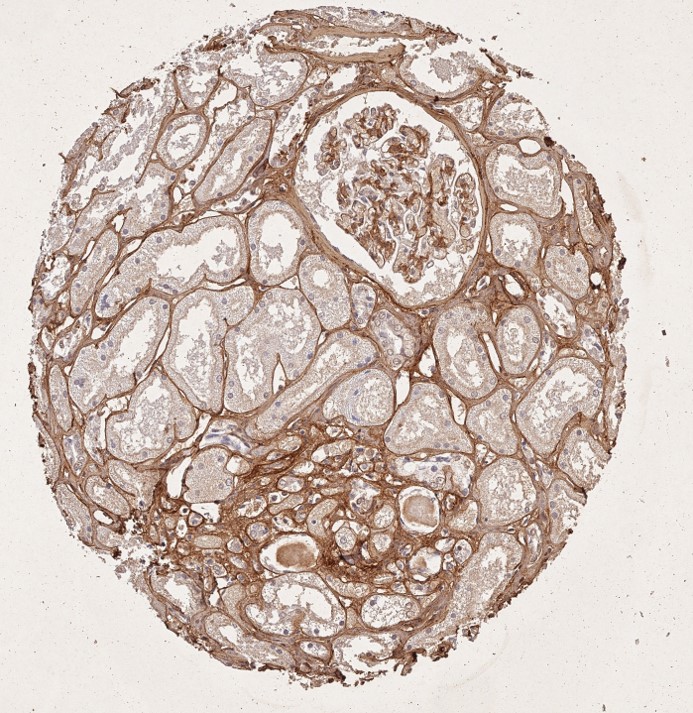
Immunohistochemistry results of Rabbit Anti-Collagen Type I Antibody. Tissue: human tubuli and blood vessels. Fixation: FFPE. Antigen Retrieval: HIER using Tris-EDTA-citrate buffer pH 7.8 for 5 min. Blocking: Peroxidase-Blocking Solution for 10 min. Primary Antibody: Anti-Collagen Type I (p/n 600-401-103-0.1) at 1:15 for 1 hr at 37 °C. Secondary Antibody: Dako REAL EnVision Detection Kit, Polymer-HRP, Rabbit/Mouse. Counterstain: Hematoxylin for 15 sec.Substrate: DAB-Chromogen, Rabbit/Mouse. Staining/Results: Intense collagen I staining of fibres surrounding tubuli and around blood vessels. Independently Validated by?antibodies-online GmbH (p/n ABIN7565871/ ABIN5596819/ ABIN5596820) by?MS Validated Antibodies.
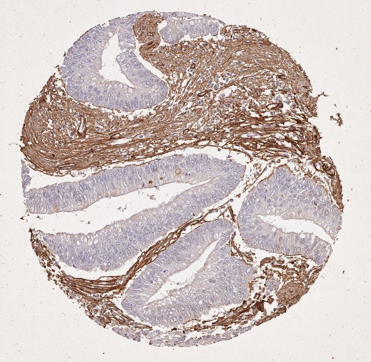
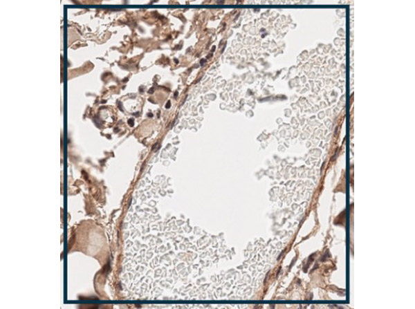
Immunohistochemistry results of Rabbit Anti-Collagen Type I Antibody. Tissue: human stroma of a colorectal adenocarcinoma.Fixation: FFPE. Antigen Retrieval: HIER using Tris-EDTA-citrate buffer pH 7.8 for 5 min. Blocking: Peroxidase-Blocking Solution for 10 min. Primary Antibody: Anti-Collagen Type I (p/n 600-401-103-0.1) at 1:15 for 1 hr at 37 °C. Secondary Antibody: Dako REAL EnVision Detection Kit, Polymer-HRP, Rabbit/Mouse. Counterstain: Hematoxylin for 15 sec.Substrate: DAB-Chromogen, Rabbit/Mouse. Staining/Results: Cancer cells are collagen I negative. Independently Validated by?antibodies-online GmbH (p/n ABIN7565871/ ABIN5596819/ ABIN5596820) by?MS Validated Antibodies.
Immunohistochemistry using Rabbit Anti-Collagen Type I Antibody. Tissue: Human Skin. Fixation: formalin fixed paraffin embedded. Antigen retrieval: HEIR pH 6.0 buffer for 20 mins. Primary antibody: Anti-Collagen Type I Antibody (lot 52110) at 1:500 for 30 mins. Secondary antibody: Anti-Rabbit Poly-HRP IgG for 8 mins. Staining: DAB kit. Localization: Peri-follicular and dermal collagen. Size: W 164.6μm x L 206.2μm.
Immunohistochemistry using Rabbit Anti-Collagen Type I Antibody. Tissue: Human Skin. Fixation: formalin fixed paraffin embedded. Primary antibody: Anti-Collagen Type I Antibody (lot 52110) at 1:500 for 30 mins. Secondary antibody: Anti-Rabbit Poly-HRP IgG for 8 mins. Antigen retrieval: HEIR pH 6.0 buffer for 20 mins. Staining: DAB kit. Localization: Vessel and dermal Collagen. Size: W 193.0μm x L 202.7μm.
|
|
|
|
Rockland's Affinity Purified anti-Collagen I antibody was used at a 1:100 dilution to detect distal tubules in normal kidney tissue. Note the absence of staining of glomeruli. The antibody was reacted with antibody for 4 hours at room temperature followed by the addition of secondary antibody and substrate reaction. Tissue was formalin-fixed and paraffin embedded. No antigen retrieval was performed.
|
|
| 別品名 |
rabbit anti-collagen type I antibody, Collagen Of Skin Tendon And Bone, Collagen Type 1 antibody, Collagen type I alpha 1 antibody, Collagen alpha-1 (I) chain, Alpha-1 type I collagen, type 1 procollagen alpha 1
|
| 由来詳細 |
[Species]Human/Bovine
|
| 交差種 |
Human
Mouse
Rat
Bovine
Porcine
|
| 適用 |
Western Blot
Immunohistochemistry
Dot Blot
|
| 免疫動物 |
Rabbit
|
| 標識物 |
Unlabeled
|
| 精製度 |
Affinity Purified
|
| GENE ID |
1277
|
| Accession No.(Gene/Protein) |
NP_000079.2, P02452
|
| Gene Symbol |
COL1A1/A2
|
| 参考文献 |
[Pub Med ID]39980135
|
| [注意事項] |
濃度はロットによって異なる可能性があります。メーカーDS及びCoAからご確認ください。
|
|
| メーカー |
品番 |
包装 |
|
RKL
|
600-401-103S
|
25 UL
|
※表示価格について
|
※当社では商品情報の適切な管理に努めておりますが、表示される法規制情報は最新でない可能性があります。
また法規制情報の表示が無いものは、必ずしも法規制に非該当であることを示すものではありません。
商品のお届け前に最新の製品法規制情報をお求めの際はこちらへお問い合わせください。
|
※当社取り扱いの試薬・機器製品および受託サービス・創薬支援サービス(納品物、解析データ等)は、研究用としてのみ販売しております。
人や動物の医療用・臨床診断用・食品用としては、使用しないように、十分ご注意ください。
法規制欄に体外診断用医薬品と記載のものは除きます。
|
|
※リンク先での文献等のダウンロードに際しましては、掲載元の規約遵守をお願いします。
|
|
※CAS Registry Numbers have not been verified by CAS and may be inaccurate.
|
