
| 別品名 |
Ido, Indo, Indoleamine 2,3-dioxygenase 1, Indoleamine-pyrrole 2,3-dioxygenase, Ido 1, Ido-1, IDO1 antibody, anti-IDO1 antibody
|
| 種由来 |
Mouse
|
| 標識物 |
Unlabeled
|
| 精製度 |
Ig fraction - Protein G
|
| 適用 |
Western Blot
Enzyme Linked Immunosorbent Assay
Immunohistochemistry
Immuno Fluorescence
Flow Cytometry
|
| 免疫動物 |
Mouse
|
| 抗体クラス |
IgG1
|
| クローン |
2E2.6
|
| 交差種 |
Mouse
|
| GENE ID |
15930
|
| Accession No.(Gene/Protein) |
NP_032350, P28776
|
| Gene Symbol |
Ido1
|
| 形状 |
滅菌済み液状品
|
| 参考文献 |
Mellor AL., et al. (2004) Nat Rev Immunol 4:762-774. Godin-Ethier J., et al. (2011) Clin Cancer Res 17:6985-6991. Mellor AL., et al. (2002) J Immunol 168:3771-3776. Frumento G., et al. (2002) J Exp Med 196:459-468. Muller AJ., et al. (2005) Nat Med 11:312-319. Lob S., et al. (2009) Nat Rev Cancer 9:445-452. Uyttenhove C., et al. (2003) Nat Med 9:1269-1274. Daubener w., et al. (1999) Adv Exp Med Biol 467:517-524. Wechsler-Reya, R., D. et al. (1997) J. Biol Chem 272:31453-31458.
|
| [注意事項] |
濃度はロットによって異なる可能性があります。メーカーDS及びCoAからご確認ください。
|
|
※サムネイル画像をクリックすると拡大画像が表示されます。
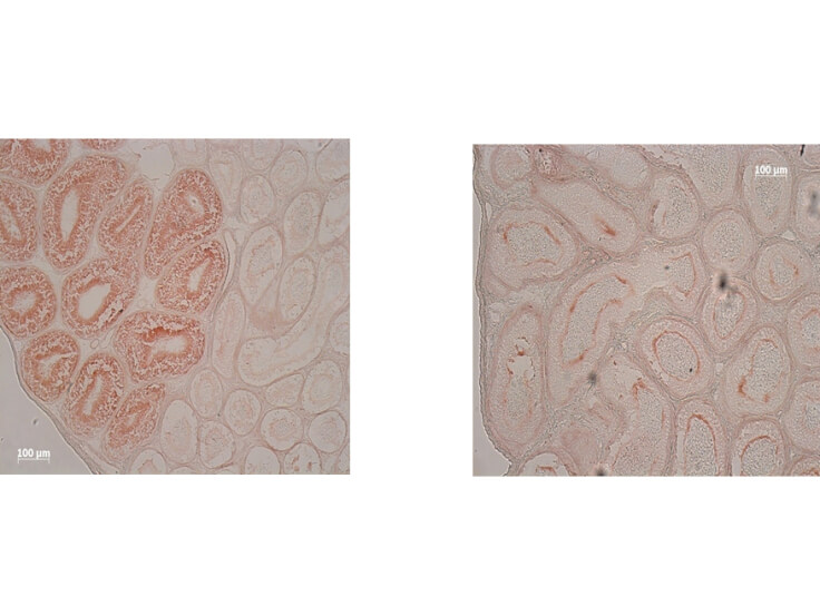
Immunohistochemistry of Mouse Anti IDO1 Antibody. Tissue: epididymis from wild type (left) or IDO1 null mice (right). Fixation: frozen sections. Antigen retrieval: not required. Primary antibody: IDO1 (2E2) monoclonal antibody. Secondary antibody: Peroxidase mouse secondary antibody at 1:10,000 for 45 min at RT. Localization: IDO 1 is located in the cytosol. Staining: IDO 1 as precipitated brown signal.
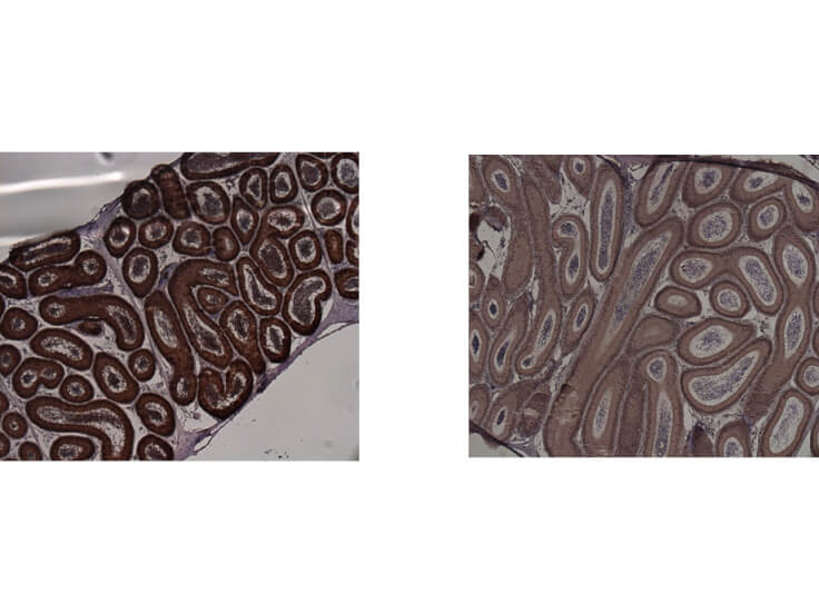
Immunohistochemistry of Mouse anti-IDO1 antibody. Tissue: epididymis from wild-type (left) or IDO1 null mice (right). Fixation: paraffin-embedded. Primary antibody: IDO1 (2E2) monoclonal antibody. Secondary antibody: Peroxidase mouse secondary antibody at 1:10,000 for 45 min at RT. Localization: IDO-1 is located in the cytosol. Staining: IDO 1 as precipitated brown signal.
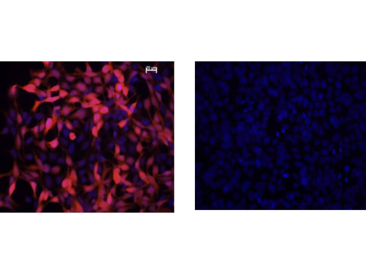
Immunofluorescence Microscopy of Mouse Anti-IDO1 Antibody. Cells: HEK293 cells. Fixation: 0.5% PFA. Expressing: mouse IDO-1 (left) and mouse IDO-2 (right). Primary antibody: IDO1 (2E2) monoclonal antibody. Antigen retrieval: not required. Secondary antibody: mouse secondary antibody at 1:10,000 for 45 min at RT. Localization: IDO-1 is located in the cytosol. Staining: IDO1 as red fluorescent signal with bis-benzimide nuclear counterstain (blue).
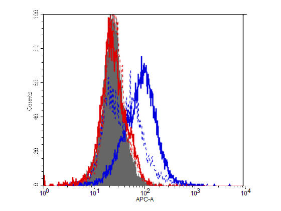
Flow Cytometry of Mouse Anti-IDO1 antibody. Cells: HEK293 cells. Expresing: mouse IDO-1(blue) and mouse IDO-2 (red). Primary antibody: IDO1 (2E2) monoclonal antibody. Secondary antibody: Biotin mouse secondary antibody at 1:10,000 for 45 min at RT and streptavidin PE at 1:5,000 for 30 min at RT.
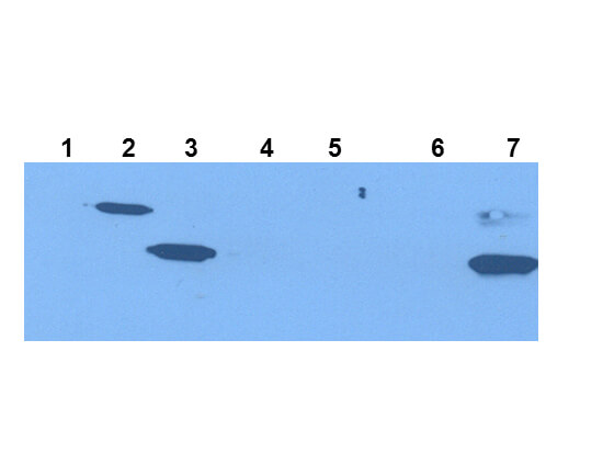
Western Blot of Mouse Anti-IDO1 Antibody. Extracts from 293HEK Cells expressing: Lane 1: Control Vector. Lane 2: His-tagged mouse IDO1. Lane 3: mouse IDO1. Lane 4: His-tagged mouse IDO2. Lane 5: mouse IDO2. Lane 6: Epididymis from IDO null. Lane 7: wild type mice. Primary antibody: IDO-1(2E2) monoclonal antibody. Secondary antibody: IRDye800TM mouse secondary antibody at 1:10,000 for 45 min at RT. Block: 1xPBST overnight at 4C. Predicted/Observed size: 41-42 kDa/41-42 kDa for IDO-1. Other band(s): none.
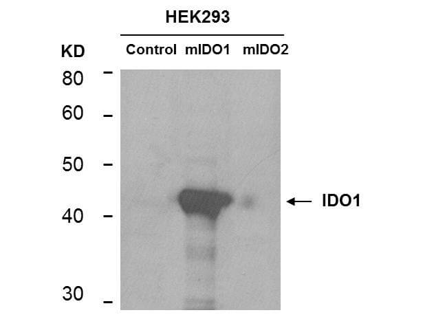
Western Blot of mouse anti-IDO1 antibody. Lane 1: HEK293 control vector. Lane 2: HEK293 expressing mouse IDO1. Lane 3: HEK293 expressing mouse IDO2. Load: 35 ug per lane. Primary antibody: IDO 1 antibody at 1:400 for overnight at 4C. Secondary antibody: IRDye800TM mouse secondary antibody at 1:10,000 for 45 min at RT. Block: 5% BLOTTO overnight at 4C. Predicted/Observed size: 45.6 kDa, ~44 kDa for IDO1. Other band(s): non-specifics.
|
|
|
|
Immunohistochemistry of Mouse Anti IDO1 Antibody. Tissue: epididymis from wild type (left) or IDO1 null mice (right). Fixation: frozen sections. Antigen retrieval: not required. Primary antibody: IDO1 (2E2) monoclonal antibody. Secondary antibody: Peroxidase mouse secondary antibody at 1:10,000 for 45 min at RT. Localization: IDO 1 is located in the cytosol. Staining: IDO 1 as precipitated brown signal.
|
|
|
| メーカー |
品番 |
包装 |
|
RKL
|
210-301-E58
|
100 UG
|
※表示価格について
| 当社在庫 |
なし
|
| 納期目安 |
約10日
|
| 保存温度 |
-20℃
|
|

