|
※サムネイル画像をクリックすると拡大画像が表示されます。
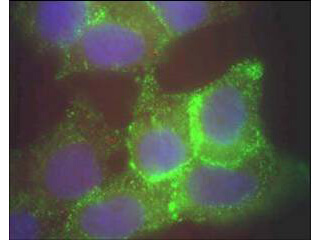
Immunofluorescence microscopy using Rockland's Protein A purified anti-Jagged-1 antibody of human corneal epithelial cells.? Primary antibody was used at a 1:500 dilution. The Jagged1 (green staining) is localized to the cytoplasm and is consistent with reports in the literature. The nucleus is stained with Bis benzamine (blue).? Personal Communication.? Aihua Ma, Univdersity of Cardiff.
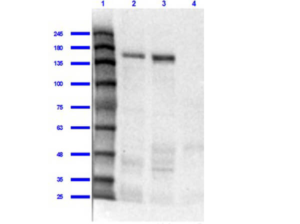
Western Blot of Rabbit Anti-Jagged 1 Antibody. Lane 1: Opal Prestained MW marker (p/n MB-210-0500). Lane 2: Mouse Liver Whole Cell Lysate [10μg] (p/n W10-000-T020). Lane 3: Human Liver Whole Cell Lysate [10μg]. Lane 4: Human Lung Whole Cell Lysate [10μg]. Primary Antibody: Anti-Jagged 1 at 1:1000 overnight at 2-8°C. Secondary Antibody: Goat Anti-Rabbit IgG Peroxidase Conjugated (p/n 611-103-122) at 1:70000 for 30mins at RT. Blocking Buffer: BlockOut Buffer (p/n MB-073) for 1hr RT. Predicted Molecular Weight: 113kDa. Exposure: 30 sec.
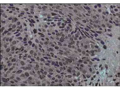
Immunohistochemical staining of human cervical cancer tissue (40X magnification) using Rockland's Protein A purified anti-Jagged-1 antibody. Tissue was fixed with formalin and embedded in paraffin. Hematoxylin was used to counter-stain cells. A 1:100 dilution of primary antibody was used. Personal Communication. Martin Kast Laboratory.
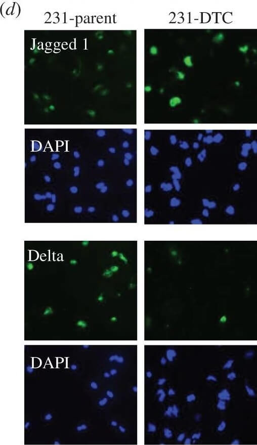
(a) Representative confocal microscopy shows CD44, CD24 and cleaved notch (NICD) in a population of drug naive MDA-MB-231. Yellow arrows indicate CD44HiCD24Lo (M) population of cells and the white arrows indicate the CD44HiCD24Hi (E/M) cells. Histogram (right panel) shows quantification of NICD in the distinct phenotype populations (M versus E/M). N = 3 biological replicates. (b) Schematic describes the experimental protocol to generate drug-tolerant cells (DTCs) parental MDA-MB-231 cells were treated with docetaxel at 100 nM (20× the IC50) and subsequently selected by substrate re-attachment and acute population outgrowth. (c) Representative confocal microscopy shows CD44, CD24 and NICD in the MDA-MB-231 parent and DTC populations. Right panel shows quantification of fluorescence intensity of each signal determined by at least 25 individual fields. N = 3 biological replicates. (d) Representative confocal microscopy shows Jagged and Delta expression in MDA-MB-231 parent and DTC. DAPI nuclear stain (blue). N = 3 biological replicates. Figure provided by CiteAb. Source: J R Soc Interface, PMID: 27170649.
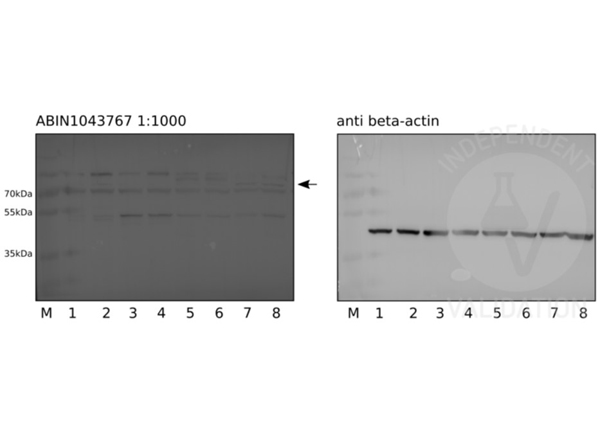
Western blot Anti-Jagged-1 (RABBIT) Antibody. Human pluripotent stem cells (lanes 1 and 2), definite endoderm cells (lanes 3 and 4), pancreatic endoderm cells (lanes 5 and 6), and pancreatic progenitors (lanes 7 and 8). Primary Antibody: Anti-Jagged-1 used at a 1:1000 (30sec exposure) overnight at 4°C. Beta-actin served as loading control at 1:2000 at RT for 1hr. ?Secondary Antibody: donkey anti-rabbit HRP conjugated antibody or donkey anti-mouse HRP conjugated antibody diluted 1:5000 for 1h at RT. Independently Validated by?antibodies-online GmbH (p/n ABIN1043767/ ABIN129524) courtesy of? Ulm University Hospital.
|
|
|
|
Immunofluorescence microscopy using Rockland's Protein A purified anti-Jagged-1 antibody of human corneal epithelial cells.? Primary antibody was used at a 1:500 dilution. The Jagged1 (green staining) is localized to the cytoplasm and is consistent with reports in the literature. The nucleus is stained with Bis benzamine (blue).? Personal Communication.? Aihua Ma, Univdersity of Cardiff.
|
|
| 別品名 |
rabbit anti-Jagged 1 Antibody, rabbit anti-Jagged1 Antibody, rabbit anti-Jagged-1 Antibody, Ser 1 antibody, AGS antibody, AHD antibody, AWS antibody, CD 339 antibody, CD339 antibody, CD339 antigen antibody, Headturner antibody, HJ1 antibody, Htu antibody
|
| 交差種 |
Human
Mouse
|
| 適用 |
Western Blot
Enzyme Linked Immunosorbent Assay
Immunohistochemistry
Immuno Fluorescence
|
| 免疫動物 |
Rabbit
|
| 抗原部位 |
a.a.110-125
|
| 標識物 |
Unlabeled
|
| 精製度 |
Affinity Purified
|
| GENE ID |
182
|
| Accession No.(Gene/Protein) |
4557679, P78504
|
| Gene Symbol |
JAG1
|
| 参考文献 |
[Pub Med ID]27170649
|
| [注意事項] |
濃度はロットによって異なる可能性があります。メーカーDS及びCoAからご確認ください。
|
|
| メーカー |
品番 |
包装 |
|
RKL
|
200-401-698S
|
25 UL
|
※表示価格について
| 当社在庫 |
なし
|
| 納期目安 |
約10日
|
| 保存温度 |
-20℃
|
|
※当社では商品情報の適切な管理に努めておりますが、表示される法規制情報は最新でない可能性があります。
また法規制情報の表示が無いものは、必ずしも法規制に非該当であることを示すものではありません。
商品のお届け前に最新の製品法規制情報をお求めの際はこちらへお問い合わせください。
|
※当社取り扱いの試薬・機器製品および受託サービス・創薬支援サービス(納品物、解析データ等)は、研究用としてのみ販売しております。
人や動物の医療用・臨床診断用・食品用としては、使用しないように、十分ご注意ください。
法規制欄に体外診断用医薬品と記載のものは除きます。
|
|
※リンク先での文献等のダウンロードに際しましては、掲載元の規約遵守をお願いします。
|
|
※CAS Registry Numbers have not been verified by CAS and may be inaccurate.
|
