
| 別品名 |
RAC-PK-alpha, Protein kinase B, PKB, C-AKT, RAC-alpha serine/threonine-protein kinase, Proto-oncogene c-Akt, AKT1, AKT 1, AKT-1, AT594, ATTO 594, ATTO-TEC 594
|
| 種由来 |
Human
|
| 標識物 |
ATTO 594
|
| 精製度 |
Ig fraction - Protein A
|
| 適用 |
Western Blot
|
| 免疫動物 |
Mouse
|
| 抗体クラス |
IgG1κ
|
| クローン |
17F6.B11
|
| 交差種 |
Human
Mouse
Rat
Monkey
|
| 翻訳後修飾 |
リン酸化
|
| GENE ID |
207
|
| Accession No.(Gene/Protein) |
P31749
|
| Gene Symbol |
AKT1
|
| 形状 |
凍結乾燥品
|
| 参考文献 |
Lawlor, M. A. and Alessi, D.R. (2001). PKB/AKT: a key mediator of cell proliferation, survival and insulin responses. J. Cell Science 114:2903-2910. Alessi, D. R. (2001). Discovery of PDK1, one of the missing links in insulin signal transduction. Biochem. Soc. Trans. 29,1 -14. Jones,P.F., Jakubowicz,T., Pitossi,F.J., Maurer,F. and Hemmings,B.A. (1991) Molecular cloning and identification of a serine/threonine protein kinase of the second-messenger subfamily. Proc. Natl. Acad. Sci. U.S.A. 88 (10), 4171-4175.
|
| [注意事項] |
濃度はロットによって異なる可能性があります。メーカーDS及びCoAからご確認ください。
|
|
※サムネイル画像をクリックすると拡大画像が表示されます。
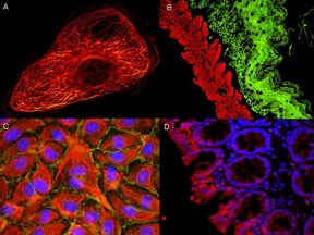
ATTO dyes can be used for multicolor immunofluorescent detection with low background and high signal. Examples shown are: A. Tubulin in PtK2 male Rat Kangaroo Kidney Epithelial Cells was detected using ATTO 532 labeled secondary antibody. B. Muscle alpha actin was stained with a mouse primary antibody and ATTO 488 anti mouse IgG (green) while Cytokeratin was stained with polyclonal rabbit anti cytokeratin and ATTO 647N anti rabbit IgG (red). C. HUVEC (Human umbilical vein endothelial cells were stained with anti Vimentin ATTO 532 (green), anti E Cadherin ATTO 655 (red) and DAPI (blue). D. Rat colon sections were stained with Anti Aquaporin 3 ATTO 594 antibody. Hoechst 33342 (blue) is used as counterstain.
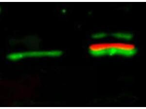
Western Blot of Mouse Anti-Akt pS473 antibody. Lane 1: unstimulated NIH/3T3 lysates contain inactive unphosphorylated Akt1, green band. Lane 2: PDGF stimulated NIH/3T3 lysate contains both inactive (green band) and activated phosphorylated Akt1 (red band). Load: 10 μg per lane. Primary antibody: rabbit anti-Akt (pan) and mouse anti-Akt pS473 specific antibodies at 1:400 for overnight at 4°C. Secondary antibody: DyLight? 549 conjugated anti-rabbit IgG (green) and DyLight? 649 conjugated anti-mouse IgG (red) secondary antibodies at 1:10,000 for 45 min at RT. Block: 5% BLOTTO overnight at 4°C.
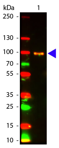
Western Blot of Mouse anti-AKT pS473 antibody Atto 594 Conjugated. Lane 1: GST Tagged AKT 1 Active Recombinant Protein. Lane 2: None. Load: 25 ng per lane. Primary antibody: None. Secondary antibody: Atto 594 mouse secondary antibody at 1:1,000 for 60 min at RT. Block: MB-070 for 30 min at RT. Predicted/Observed size: ~100 kDa, ~100 kDa for AKT pS473. Other band(s): None
|
|
|
|
ATTO dyes can be used for multicolor immunofluorescent detection with low background and high signal. Examples shown are: A. Tubulin in PtK2 male Rat Kangaroo Kidney Epithelial Cells was detected using ATTO 532 labeled secondary antibody. B. Muscle alpha actin was stained with a mouse primary antibody and ATTO 488 anti mouse IgG (green) while Cytokeratin was stained with polyclonal rabbit anti cytokeratin and ATTO 647N anti rabbit IgG (red). C. HUVEC (Human umbilical vein endothelial cells were stained with anti Vimentin ATTO 532 (green), anti E Cadherin ATTO 655 (red) and DAPI (blue). D. Rat colon sections were stained with Anti Aquaporin 3 ATTO 594 antibody. Hoechst 33342 (blue) is used as counterstain.
|
|
|
| メーカー |
品番 |
包装 |
|
RKL
|
200-355-268
|
100 UG
|
※表示価格について
| 当社在庫 |
なし
|
| 納期目安 |
約10日
|
| 保存温度 |
4℃
|
|

