
| 別品名 |
phospho AKT, RAC-PK-alpha, Protein kinase B, PKB, C-AKT, RAC-alpha serine/threonine-protein kinase, Proto-oncogene c-Akt, AKT1, AKT 1, AKT-1, phospho AKT antibody, Anti-AKT pS473 Antibody Biotin Conjugated
|
| 種由来 |
Human
|
| 標識物 |
Biotin
|
| 精製度 |
Ig fraction - Protein A
|
| 適用 |
Western Blot
Enzyme Linked Immunosorbent Assay
Immunohistochemistry
|
| 免疫動物 |
Mouse
|
| 抗体クラス |
IgG1κ
|
| クローン |
17F6.B11
|
| 交差種 |
Human
Mouse
Rat
Monkey
|
| 翻訳後修飾 |
リン酸化
|
| GENE ID |
207
|
| Accession No.(Gene/Protein) |
P31749
|
| Gene Symbol |
AKT1
|
| 形状 |
凍結乾燥品
|
| 参考文献 |
Lawlor, M. A. and Alessi, D.R. (2001). PKB/AKT: a key mediator of cell proliferation, survival and insulin responses. J. Cell Science 114:2903-2910. Alessi, D. R. (2001). Discovery of PDK1, one of the missing links in insulin signal transduction. Biochem. Soc. Trans. 29,1 -14. Jones,P.F., Jakubowicz,T., Pitossi,F.J., Maurer,F. and Hemmings,B.A. (1991) Molecular cloning and identification of a serine/threonine protein kinase of the second-messenger subfamily. Proc. Natl. Acad. Sci. U.S.A. 88 (10), 4171-4175.
|
| [注意事項] |
濃度はロットによって異なる可能性があります。メーカーDS及びCoAからご確認ください。
|
|
※サムネイル画像をクリックすると拡大画像が表示されます。
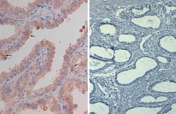
Immunohistochemistry of mouse Anti AKT pS473 (MOUSE) Biotin Conjugated. Tissue: prostate at 40X. Fixation: FFPE buffered formalin 10% conc. Antigen retrieval: Heat, Citrate pH 6.2. Pressure Cooker, left. (pH 9 shown on right as negative control). Primary antibody: AKTsS473 biotin 20 ug/mL for 1 h at RT. Secondary antibody: Streptavidin Conj. HRP at 10 ug/ml. Localization: nuclear and occasionally cytoplasmic. Staining: antibody as precipitated red signal with a hematoxylin purple nuclear counterstain.
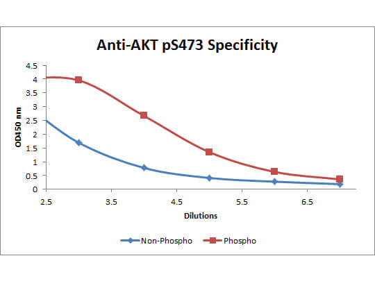
ELISA of Mouse anti-Akt phospho S473 Biotin Conjugated antibody. Antigen: BSA conjugates of Akt phospho S473 and AKT non-phospho S473. Coating amount: 0.1 ug per well. Primary antibody: Akt phospho S473 Biotin Conjugated antibody at 5 ug/mL. Dilution series: 3-fold. Mid-point concentration: 5 ng/mL Akt phospho S473 Biotin Conjugated antibody. Secondary antibody: Peroxidase streptavidin secondary antibody at 1:10,000. Substrate: TMB (p/n TMBE-0100)
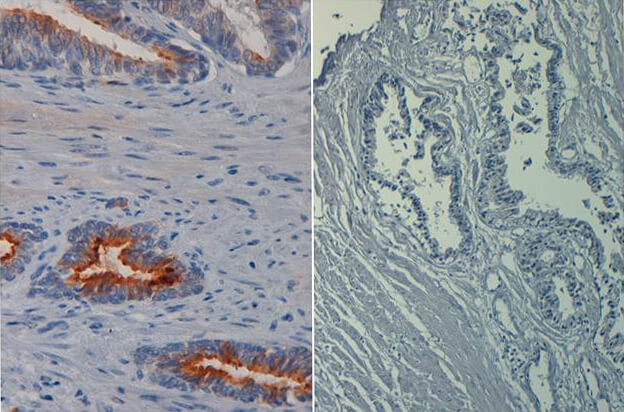
Immunohistochemistry of mouse anti AKT phospho S473 biotin conjugated. Tissue: prostate at 40X (left) with negative control (right). Fixation: FFPE buffered formalin 10% conc. Antigen retrieval: Heat, Citrate pH 6.2. Pressure Cooker. Primary antibody: AKT pS473 biotin at 20 ug/mL for 1 h at RT. Secondary antibody: Streptavidin Conj. HRP at 10 ug/ml. Localization: nuclear and occasionally cytoplasmic. Staining: antibody as precipitated red signal with a hematoxylin purple nuclear counterstain.
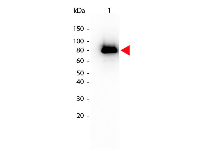
Western Blot of Mouse anti-Akt phospho S473 Biotin Conjugated antibody. Lane 1: GST tagged AKT1 active recombinant protein. Lane 2: none. Load: 25 ng per lane. Primary antibody: Akt phospho S473 Biotin Conjugated antibody at 1:1,000 for overnight at 4C. Secondary antibody: HRP Streptavidin secondary antibody at 1:40,000 for 30 min at RT. Block: MB-070 for 30 min at RT. Predicted/Observed size: 79 kDa, 79 kDa for Akt phospho S473. Other band(s): none
|
|
|
|
Immunohistochemistry of mouse Anti AKT pS473 (MOUSE) Biotin Conjugated. Tissue: prostate at 40X. Fixation: FFPE buffered formalin 10% conc. Antigen retrieval: Heat, Citrate pH 6.2. Pressure Cooker, left. (pH 9 shown on right as negative control). Primary antibody: AKTsS473 biotin 20 ug/mL for 1 h at RT. Secondary antibody: Streptavidin Conj. HRP at 10 ug/ml. Localization: nuclear and occasionally cytoplasmic. Staining: antibody as precipitated red signal with a hematoxylin purple nuclear counterstain.
|
|
|
| メーカー |
品番 |
包装 |
|
RKL
|
200-306-268
|
50 UG
|
※表示価格について
| 当社在庫 |
なし
|
| 納期目安 |
約10日
|
| 保存温度 |
4℃
|
|

