|
※サムネイル画像をクリックすると拡大画像が表示されます。
Figure 1. Western Blot testing of anti-CDNF polyclonal antibody.
Line 1. PageRuler Prestained Protein Ladder (#SM0671 Fermentas);
Line 2. Recombinant CDNF expressed into the supernatant of CHO cell culture medium.

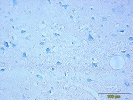
Figure 2. Immunohistochemistry testing of anti-CDNF rabbit polyclonal antibody.
Analysis was performed using formalin-fixed paraffin-embedded human cerebral cortex tissue sections from Alzheimer’s disease patients. Tissue sections were boiled with sodium citrate buffer (pH 6) for antigen retrieval. Incubation with primary antibody at 5 μg/ml was performed overnight at 4°C. DAKO EnVisionTM Detection System, Peroxidase/DAB was used for visualization. Sections were counterstained with toluidine blue and mounted with Eukitt mounting medium. A. CDNF staining by rabbit polyclonal anti-CDNF antibody; B. Negative staining without primary antibody

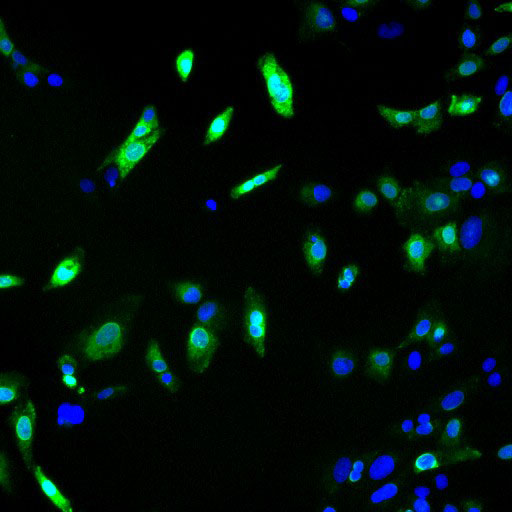
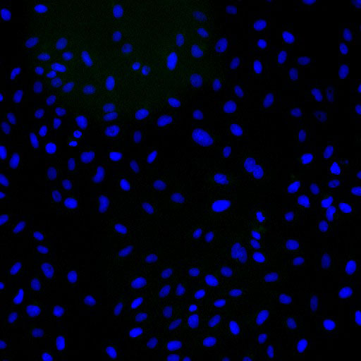
Figure 3. Immunofluorescence detection of human CDNF expressed in U2OS cells.
CDNF was visualized using anti-CDNF rabbit polyclonal antibody, dilution 1:3000. Goat ant-rabbit AlexaFluor488 was used as secondary antibody. For nuclear staining DAPI was used. ArrayScan VTI platform (Thermo Scientific) was used for image acquisition (10x objective). Composite picture was generated using pseudocolors green for CDNF specific signal and blue for nuclei. A. CDNF-expressing U2OS cells; B. Negative control (non-transfected U2OS cells)
|
|
|
|
Figure 1. Western Blot testing of anti-CDNF polyclonal antibody.
Line 1. PageRuler Prestained Protein Ladder (#SM0671 Fermentas);
Line 2. Recombinant CDNF expressed into the supernatant of CHO cell culture medium.
|
|
| 種由来 |
Human
|
| 交差種 |
Human
|
| 適用 |
Western Blot
IHC paraffin embedding section
Enzyme Linked Immunosorbent Assay
Immuno Fluorescence
|
| 免疫動物 |
Rabbit
|
| 抗体クラス |
IgG
|
|
| メーカー |
品番 |
包装 |
|
QTM
|
300-100
|
100 UG
|
※表示価格について
|
※当社では商品情報の適切な管理に努めておりますが、表示される法規制情報は最新でない可能性があります。
また法規制情報の表示が無いものは、必ずしも法規制に非該当であることを示すものではありません。
商品のお届け前に最新の製品法規制情報をお求めの際はこちらへお問い合わせください。
|
※当社取り扱いの試薬・機器製品および受託サービス・創薬支援サービス(納品物、解析データ等)は、研究用としてのみ販売しております。
人や動物の医療用・臨床診断用・食品用としては、使用しないように、十分ご注意ください。
法規制欄に体外診断用医薬品と記載のものは除きます。
|
|
※リンク先での文献等のダウンロードに際しましては、掲載元の規約遵守をお願いします。
|
|
※CAS Registry Numbers have not been verified by CAS and may be inaccurate.
|