|
※サムネイル画像をクリックすると拡大画像が表示されます。
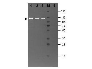
Western blotting using Rockland's Fluorescein conjugated anti-b-Galactosidase antibody shows a band at ~117 kDa (lanes 1 - 3) corresponding to 60 ng, 30 ng and 15 ng, respectively of b-Gal present in partially purified preparations (arrowhead). Lane 4 shows no cross reactivity with proteins present in a non-specific control E.coli lysate. Proteins were resolved on a 4-20% Tris-Glycine gel by SDS-PAGE and transferred to nitrocellulose and blocking using Blocking Buffer for Fluorescent Western Blotting (p/n MB-070). The membrane was probed with fluorescein conjugated anti-b-Galactosidase (p/n 200-4236) diluted to 1:10,000. Reaction occurred for 2 hours at room temperature. Molecular weight estimation was made by comparison to a prestained MW marker in lane M. Fluorescence image was captured using the VersaDocR Imaging System developed by BIO-RAD. Other detection systems will yield similar results
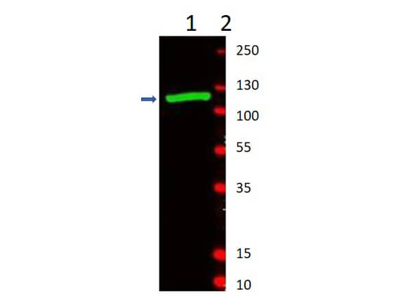
Western Blot of Rabbit Anti-Beta-Galactosidase Antibody. Lane 1: partially purified preparation b-Galactosidase [1.0μg]. Lane 2: Molecular Weight Marker. Primary Antibody: Anti-Beta-Galactosidase at 1:1000 overnight at 2-8°C. Secondary Antibody: Goat Anti-Rabbit IgG IRDyeR800 (p/n 611-132-122) 1:10,000 for 45min at RT. Observed MW: ~117kDa.
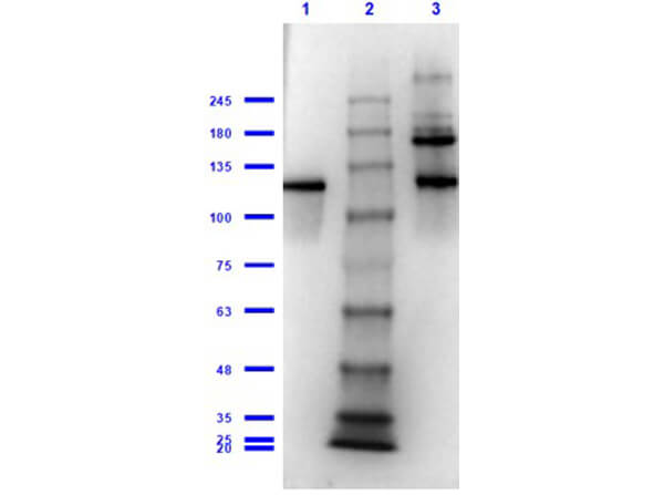
Western Blot of Rabbit Anti-Beta-Galactosidase Antibody. Lane 1: Beta-Galactosidase Reduced [0.1μg]. Lane 2: Opal Prestained Molecular Weight Marker (p/n MB-210-0500). Lane 3: Beta-Galactosidase Non-Reduced [0.1μg]. Primary Antibody: Anti-Beta-Galactosidase at 1:1000 overnight at 2-8°C. Secondary Antibody: Goat Anti-Rabbit IgG HRP (p/n 611-103-122) 1:70,000 for 30min at RT. Expected MW: ~117kDa.
|
|
|
|
Western blotting using Rockland's Fluorescein conjugated anti-b-Galactosidase antibody shows a band at ~117 kDa (lanes 1 - 3) corresponding to 60 ng, 30 ng and 15 ng, respectively of b-Gal present in partially purified preparations (arrowhead). Lane 4 shows no cross reactivity with proteins present in a non-specific control E.coli lysate. Proteins were resolved on a 4-20% Tris-Glycine gel by SDS-PAGE and transferred to nitrocellulose and blocking using Blocking Buffer for Fluorescent Western Blotting (p/n MB-070). The membrane was probed with fluorescein conjugated anti-b-Galactosidase (p/n 200-4236) diluted to 1:10,000. Reaction occurred for 2 hours at room temperature. Molecular weight estimation was made by comparison to a prestained MW marker in lane M. Fluorescence image was captured using the VersaDocR Imaging System developed by BIO-RAD. Other detection systems will yield similar results
|
|
| 別品名 |
rabbit anti-Beta Galactosidase Antibody, rabbit anti-beta gal antibody, β-Gal, Anti-β-Gal Antibody
|
| 適用 |
Western Blot
Enzyme Linked Immunosorbent Assay
|
| 免疫動物 |
Rabbit
|
| 標識物 |
Unlabeled
|
| 精製度 |
Ig fraction - Ion Exchange /Gel Filtration
|
| Accession No.(Gene/Protein) |
NP_414878.1, P00722
|
| Gene Symbol |
lacZ
|
| Tag情報 |
b-GAL
|
| 参考文献 |
[Pub Med ID]14983513
|
|
| メーカー |
品番 |
包装 |
|
RKL
|
200-4136S
|
25 UL
|
※表示価格について
| 当社在庫 |
なし
|
| 納期目安 |
約10日
|
| 保存温度 |
-20℃
|
|
※当社では商品情報の適切な管理に努めておりますが、表示される法規制情報は最新でない可能性があります。
また法規制情報の表示が無いものは、必ずしも法規制に非該当であることを示すものではありません。
商品のお届け前に最新の製品法規制情報をお求めの際はこちらへお問い合わせください。
|
※当社取り扱いの試薬・機器製品および受託サービス・創薬支援サービス(納品物、解析データ等)は、研究用としてのみ販売しております。
人や動物の医療用・臨床診断用・食品用としては、使用しないように、十分ご注意ください。
法規制欄に体外診断用医薬品と記載のものは除きます。
|
|
※リンク先での文献等のダウンロードに際しましては、掲載元の規約遵守をお願いします。
|
|
※CAS Registry Numbers have not been verified by CAS and may be inaccurate.
|
