|
※サムネイル画像をクリックすると拡大画像が表示されます。
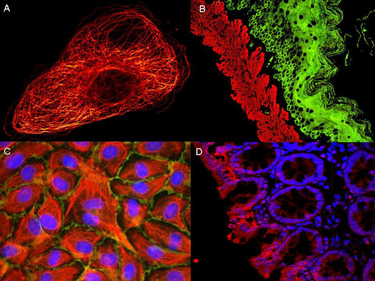
ATTO R dyes can be used for multicolor immunofluorescent detection with low background and high signal. Examples shown are: A. Tubulin in PtK2- male Rat Kangaroo Kidney Epithelial Cells was detected using ATTO 532 labeled secondary antibody. B. Muscle alpha-actin was stained with a mouse primary antibody and ATTO 488 anti-mouse IgG (green) while Cytokeratin was stained with polyclonal rabbit anti-cytokeratin and ATTO 647N anti-rabbit IgG (red). C. HUVEC (Human umbilical vein endothelial cells were stained with anti- Vimentin-ATTO 532 (green), anti-E-Cadherin-ATTO 655 (red) and DAPI (blue). D. Rat colon sections were stained with Anti-Aquaporin 3-ATTO 594 antibody. Hoechst 33342 (blue) is used as counterstain. Images provided courtesy of Dr. Jorg Reichwein, ATTO-TEC GmbH
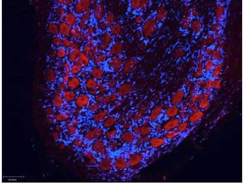
Atto? dyes can be used for multicolor immunofluorescent detection with low background and high signal. Example shown here is Immunohistochemical staining using ATTO-550 Anti-Aquaporin 2-antibody (red) of paraffin embedded region of rat kidney showing a transversal cut of the inner medulla near to the renal papilla. Nuclei are visualized with Hoechst 33342 (blue). Images provided courtesy of Dr. Jorg Reichwein, ATTO-TEC GmbH
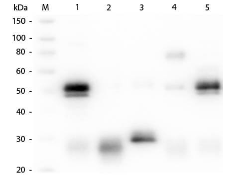
Western Blot of Unconjugated Anti-Rabbit IgG (H&L) (GOAT) Antibody (Min X Bv, Ch, Gt, GP, Ham, Hs, Hu, Ms, Rt & Sh Serum Proteins) (p/n 611-101-122). Lane M: 3 μl Molecular Ladder. Lane 1: Rabbit IgG whole molecule (p/n 011-0102). Lane 2: Rabbit IgG F(ab) Fragment (p/n 011-0105). Lane 3: Rabbit IgG F(c) Fragment (p/n 010-0103). Lane 4: Rabbit IgM Whole Molecule (p/n 011-0107). Lane 5: Normal Rabbit Serum (p/n B309). All samples were reduced. Load: 50 ng per lane. Block: MB-070 for 30 min at RT. Primary Antibody: Anti-Rabbit IgG (H&L) (GOAT) Antibody (Min X Bv, Ch, Gt, GP, Ham, Hs, Hu, Ms, Rt & Sh Serum Proteins) (p/n 611-101-122) 1:1,000 for 60 min at RT. Secondary antibody: Anti-Goat IgG (DONKEY) Peroxidase Conjugated Antibody (p/n CUST10) 1:40,000 in MB-070 for 30 min at RT. Predicted/Observed Size: 25 and 50 kDa for Rabbit IgG and Serum, 25 kDa for F(c) and F(ab), 70 and 23 kDa for IgM. Rabbit F(c) migrates slightly higher.
|
|
|
|
ATTO R dyes can be used for multicolor immunofluorescent detection with low background and high signal. Examples shown are: A. Tubulin in PtK2- male Rat Kangaroo Kidney Epithelial Cells was detected using ATTO 532 labeled secondary antibody. B. Muscle alpha-actin was stained with a mouse primary antibody and ATTO 488 anti-mouse IgG (green) while Cytokeratin was stained with polyclonal rabbit anti-cytokeratin and ATTO 647N anti-rabbit IgG (red). C. HUVEC (Human umbilical vein endothelial cells were stained with anti- Vimentin-ATTO 532 (green), anti-E-Cadherin-ATTO 655 (red) and DAPI (blue). D. Rat colon sections were stained with Anti-Aquaporin 3-ATTO 594 antibody. Hoechst 33342 (blue) is used as counterstain. Images provided courtesy of Dr. Jorg Reichwein, ATTO-TEC GmbH
|
|
| 別品名 |
Goat anti-Rabbit IgG Antibody ATTO550 Conjugation, Goat anti-Rabbit IgG ATTO 550 Conjugated Antibody
|
| 交差種 |
Rabbit
|
| 非交差(吸収処理)種 |
Human
Mouse
Rat
Bovine
Chicken
Sheep
Goat
Guinea Pig
Hamster
Equine
|
| 適用 |
Western Blot
Dot Blot
|
| 免疫動物 |
Goat
|
| 標識物 |
ATTO 550
|
| 精製度 |
Affinity Purified
|
| 参考文献 |
[Pub Med ID]30012581
|
| [注意事項] |
濃度はロットによって異なる可能性があります。メーカーDS及びCoAからご確認ください。
|
|
| メーカー |
品番 |
包装 |
|
RKL
|
611-154-122
|
500 UG
|
※表示価格について
| 当社在庫 |
なし
|
| 納期目安 |
約10日
|
| 保存温度 |
4℃
|
|
※当社では商品情報の適切な管理に努めておりますが、表示される法規制情報は最新でない可能性があります。
また法規制情報の表示が無いものは、必ずしも法規制に非該当であることを示すものではありません。
商品のお届け前に最新の製品法規制情報をお求めの際はこちらへお問い合わせください。
|
※当社取り扱いの試薬・機器製品および受託サービス・創薬支援サービス(納品物、解析データ等)は、研究用としてのみ販売しております。
人や動物の医療用・臨床診断用・食品用としては、使用しないように、十分ご注意ください。
法規制欄に体外診断用医薬品と記載のものは除きます。
|
|
※リンク先での文献等のダウンロードに際しましては、掲載元の規約遵守をお願いします。
|
|
※CAS Registry Numbers have not been verified by CAS and may be inaccurate.
|
