|
※サムネイル画像をクリックすると拡大画像が表示されます。
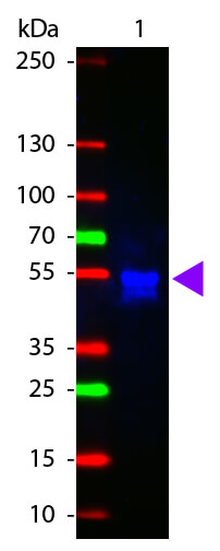
Western Blot of ATTO 425 conjugated Goat anti-Rabbit IgG antibody. Lane 1: Rabbit IgG. Lane 2: none. Load: 50 ng per lane. Primary antibody: none. Secondary antibody: ATTO 425 rabbit secondary antibody at 1:1,000 for 60 min at RT. Block: MB-070 for 30 min RT. Predicted/Observed size: 55 kDa, 28 kDa/55 kDa for Rabbit IgG. Other band(s): none.
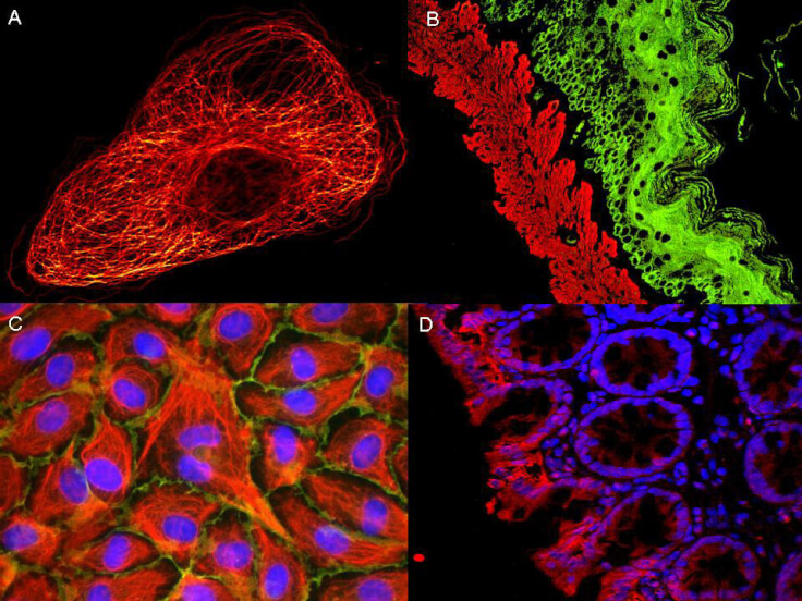
ATTO R dyes can be used for multicolor immunofluorescent detection with low background and high signal. Examples shown are: A. Tubulin in PtK2- male Rat Kangaroo Kidney Epithelial Cells was detected using ATTO 532 labeled secondary antibody. B. Muscle alpha-actin was stained with a mouse primary antibody and ATTO 488 anti-mouse IgG (green) while Cytokeratin was stained with polyclonal rabbit anti-cytokeratin and ATTO 647N anti-rabbit IgG (red). C. HUVEC (Human umbilical vein endothelial cells were stained with anti- Vimentin-ATTO 532 (green), anti-E-Cadherin-ATTO 655 (red) and DAPI (blue). D. Rat colon sections were stained with Anti-Aquaporin 3-ATTO 594 antibody. Hoechst 33342 (blue) is used as counterstain. Images provided courtesy of Dr. Jorg Reichwein, ATTO-TEC GmbH
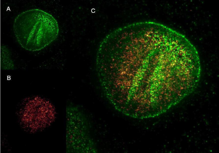
Rockland Dylight and ATTORdye conjugated antibodies provide high signal and low background for confocal microscopy (upper images) and high resolution Stimulated Emission Depletion (STED) Microscopy (lower images). Both Dylight and Atto conjugated secondary antibodies maintained robust, intense signal during repeated laser excitation and de-excitation used during STED microscopy. Shown here are: A. (Green) Mouse anti NuP (NuP=Nuclear Pore Protein) detected with Dylight 488 Goat anti mouse (610-141-121) B. (Red) Rabbit Anti Ezh1/2 Pab (Ezh=enhancer of zeste homology) with detection by Rockland ATTO R425 conjugated Goat anti Rabbit (611-151-122) (Red and Green) Images combined. Data was collected on a STED-CW TCS-SP5 Confocal system (Leica Microsystems) equipped with a DFC 350FX Camera allowing sequential acquisition in widefield, confocal and STED CW imaging modes and provided courtesy of: Myriam Gastard, PhD, personal communication, Leica Microsystems, Inc. USA
Rockland DyLight and ATTO Rdye conjugated antibodies provide high signal and low background for confocal microscopy and high resolution Stimulated Emission Depletion (STED) Microscopy. Both Dylight and Atto conjugated secondary antibodies maintained robust, intense signal during repeated laser excitation and de-excitation used during STED microscopy. Shown here are: A. (Green) Mouse anti NuP (NuP=Nuclear Pore Protein) detected with Dylight 488 Goat anti mouse (610-141-121) B. (Red) Rabbit Anti Ezh1/2 Pab (Ezh=enhancer of zeste homology) with detection by Rockland ATTO R425 conjugated Goat anti Rabbit (611-151-122) C. (Red and Green) Images combined. Data was collected on a STED-CW TCS-SP5 Confocal system (Leica Microsystems) equipped with a DFC 350FX Camera allowing sequential acquisition in wide-field, confocal and STED CW imaging modes and provided courtesy of: Myriam Gastard, PhD, personal communication, Leica Microsystems, Inc. USA
|
|
|
|
Western Blot of ATTO 425 conjugated Goat anti-Rabbit IgG antibody. Lane 1: Rabbit IgG. Lane 2: none. Load: 50 ng per lane. Primary antibody: none. Secondary antibody: ATTO 425 rabbit secondary antibody at 1:1,000 for 60 min at RT. Block: MB-070 for 30 min RT. Predicted/Observed size: 55 kDa, 28 kDa/55 kDa for Rabbit IgG. Other band(s): none.
|
|
| 別品名 |
Goat anti-Rabbit IgG Antibody ATTO425 Conjugation, Goat anti-Rabbit IgG ATTO 425 Conjugated Antibody
|
| 交差種 |
Rabbit
|
| 非交差(吸収処理)種 |
Human
Mouse
Rat
Bovine
Chicken
Sheep
Goat
Guinea Pig
Hamster
Equine
|
| 適用 |
Western Blot
Dot Blot
|
| 免疫動物 |
Goat
|
| 標識物 |
ATTO 425
|
| 精製度 |
Affinity Purified
|
| 参考文献 |
[Pub Med ID]30789343
|
| [注意事項] |
濃度はロットによって異なる可能性があります。メーカーDS及びCoAからご確認ください。
|
|
| メーカー |
品番 |
包装 |
|
RKL
|
611-151-122S
|
100 UG
|
※表示価格について
| 当社在庫 |
なし
|
| 納期目安 |
約10日
|
| 法規制 |
毒
|
| 保存温度 |
4℃
|
|
※当社では商品情報の適切な管理に努めておりますが、表示される法規制情報は最新でない可能性があります。
また法規制情報の表示が無いものは、必ずしも法規制に非該当であることを示すものではありません。
商品のお届け前に最新の製品法規制情報をお求めの際はこちらへお問い合わせください。
|
※当社取り扱いの試薬・機器製品および受託サービス・創薬支援サービス(納品物、解析データ等)は、研究用としてのみ販売しております。
人や動物の医療用・臨床診断用・食品用としては、使用しないように、十分ご注意ください。
法規制欄に体外診断用医薬品と記載のものは除きます。
|
|
※リンク先での文献等のダウンロードに際しましては、掲載元の規約遵守をお願いします。
|
|
※CAS Registry Numbers have not been verified by CAS and may be inaccurate.
|
