

| 別品名 |
AT647N, ATTO 647N, ATTO-TEC 647N
|
| 種由来 |
Goat
|
| 標識物 |
ATTO 647N
|
| 精製度 |
Affinity Purified
|
| 適用 |
Western Blot
Dot Blot
|
| 免疫動物 |
Rabbit
|
| 非交差(吸収処理)種 |
Human
Mouse
Rabbit
|
| 形状 |
凍結乾燥品
|
| [注意事項] |
濃度はロットによって異なる可能性があります。メーカーDS及びCoAからご確認ください。
|
|
※サムネイル画像をクリックすると拡大画像が表示されます。
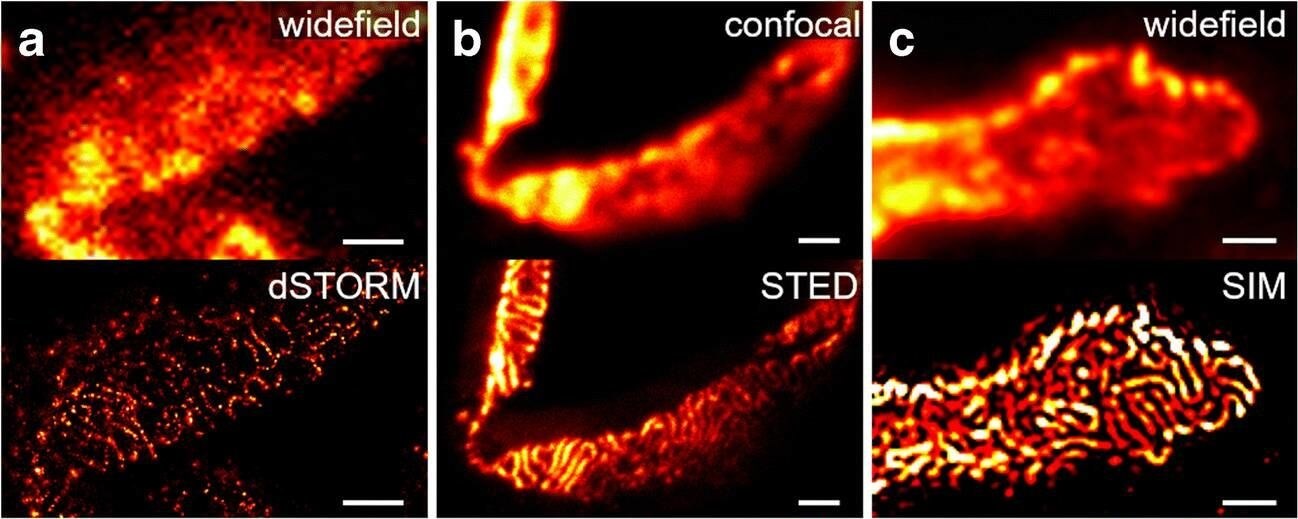
Comparison of nephrin imaging with different conventional and super resolution modalities. A) Widefield vs dSTORM using Atto647N. B) Confocal vs STED using Atto647N. C) Widefield vs SIM using Alexa Fluor 488. D) ExM prepared sample imaged with widefield vs e confocal using Alexa Fluor 488. The confocal ExM image furthermore shows the nucleus stained with Hoechst. Highlighted panels show x3 zoomed areas. Scale bars in ac are 1?μm, in d and e 10?μm and in the zoomed in areas 5?μm (after expansion) Figure provided by CiteAb. Source: Anal Bioanal Chem, PMID: 33277998.
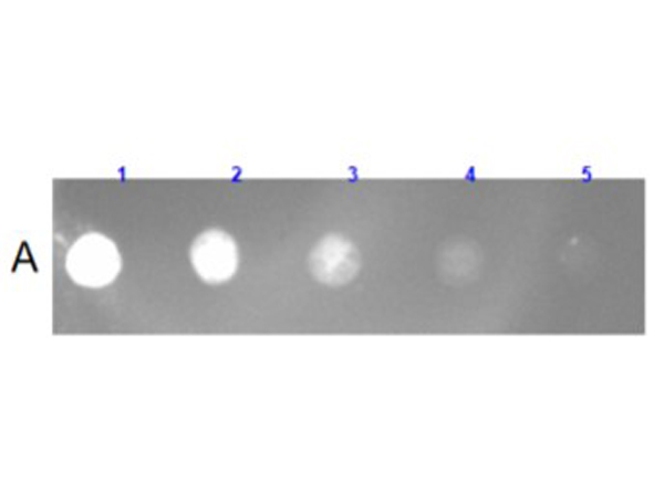
Dot Blot Results of Rabbit Anti-Goat IgG (H&L) Antibody (MX Hu, Ms, Rb) ATTO 647N Conjugated. Sample: Goat IgG [p/n 005-0102]. Loaded: 1) 100ng, 2) 33.33ng, 3) 11.11ng, 4) 3.7ng, 5) 1.23ng. Antibody: Anti-Goat IgG (H&L) [Rabbit] Antibody (MX3) ATTO 647N Conjugated at 1.0ug/mL for 1hr at RT. Block: Blocking Buffer for Fluorescent Western Blotting [p/n MB-070] for 30 mins at RT.
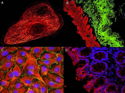
ATTO R dyes can be used for multicolor immunofluorescent detection with low background and high signal. Examples shown are: A. Tubulin in PtK2- male Rat Kangaroo Kidney Epithelial Cells was detected using ATTO 532 labeled secondary antibody. B. Muscle alpha-actin was stained with a mouse primary antibody and ATTO 488 anti-mouse IgG (green) while Cytokeratin was stained with polyclonal rabbit anti-cytokeratin and ATTO 647N anti-rabbit IgG (red). C. HUVEC (Human umbilical vein endothelial cells were stained with anti- Vimentin-ATTO 532 (green), anti-E-Cadherin-ATTO 655 (red) and DAPI (blue). D. Rat colon sections were stained with Anti-Aquaporin 3-ATTO 594 antibody. Hoechst 33342 (blue) is used as counterstain. Images provided courtesy of Dr. Jorg Reichwein, ATTO-TEC GmbH
|
|
|
|
Comparison of nephrin imaging with different conventional and super resolution modalities. A) Widefield vs dSTORM using Atto647N. B) Confocal vs STED using Atto647N. C) Widefield vs SIM using Alexa Fluor 488. D) ExM prepared sample imaged with widefield vs e confocal using Alexa Fluor 488. The confocal ExM image furthermore shows the nucleus stained with Hoechst. Highlighted panels show x3 zoomed areas. Scale bars in ac are 1?μm, in d and e 10?μm and in the zoomed in areas 5?μm (after expansion) Figure provided by CiteAb. Source: Anal Bioanal Chem, PMID: 33277998.
|
|
|
| メーカー |
品番 |
包装 |
|
RKL
|
605-456-013S
|
100 UG
|
※表示価格について
| 当社在庫 |
なし
|
| 納期目安 |
約10日
|
| 保存温度 |
4℃
|
|

