|
※サムネイル画像をクリックすると拡大画像が表示されます。
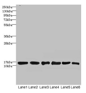
Western blot
All lanes: SOD1 antibody at 6μg/ml
Lane 1: Mouse liver tissue
Lane 2: Mouse brain tissue
Lane 3: Hela whole cell lysate
Lane 4: MCF-7 whole cell lysate
Lane 5: A549 whole cell lysate
Lane 6: HepG2 whole cell lysate
Secondary
Goat polyclonal to rabbit IgG at 1/10000 dilution
Predicted band size: 16 kDa
Observed band size: 16 kDa
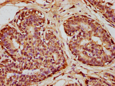
IHC image of CSB-PA02864A0Rb diluted at 1:100 and staining in paraffin-embedded human breast cancer performed on a Leica BondTM system. After dewaxing and hydration, antigen retrieval was mediated by high pressure in a citrate buffer (pH 6.0). Section was blocked with 10% normal goat serum 30min at RT. Then primary antibody (1% BSA) was incubated at 4°C overnight. The primary is detected by a biotinylated secondary antibody and visualized using an HRP conjugated SP system.
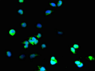
Immunofluorescent analysis of 293T cells using CSB-PA02864A0Rb at dilution of 1:100 and Alexa Fluor 488-congugated AffiniPure Goat Anti-Rabbit IgG(H+L)
|

