
| 別品名 |
Mitogen-activated protein kinase 3, ERK1, HS44KDAP, HuMKER1A, P44ERK1, P44MAPK, PRKM3, MAP kinase3, Mitogen-activated protein kinase 3
|
| 抗原部位 |
a.a.1-137
|
| 種由来 |
Human
|
| 標識物 |
Unlabeled
|
| 精製度 |
Ig fraction - Protein G
|
| 適用 |
Western Blot
Enzyme Linked Immunosorbent Assay
Immuno Fluorescence
Immunocytochemistry (cell)
|
| 免疫動物 |
Mouse
|
| 抗体クラス |
IgG2bκ
|
| クローン |
AT1A2
|
| 交差種 |
Human
|
| Accession No.(Gene/Protein) |
NP_002737.2, P27361
|
| 形状 |
液状
|
| 参考文献 |
Meloche S, et al,. Oncogene (2007) 26:3227-39
Raman M, et al,. Oncogene (2007) 26:3100-12
Roberts PJ, et al,. Oncogene (2007) 26:3291-310
Shaul YD, et al,. Seger R. Acta (2007) 1773:1213-26
|
|
※サムネイル画像をクリックすると拡大画像が表示されます。
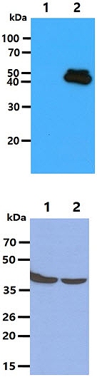
The Recombinant proteins (50ng) were resolved by SDS-PAGE, transferred to PVDF membrane and probed with anti-human MAPK3 antibody (1:500). Proteins were visualized using a goat anti-mouse secondary antibody conjugated to HRP and an ECL detection system.
Lane 1.: MAPK1 recombinant protein
Lane 2.: MAPK3 recombinant protein
The Cell lysates (40ug) were resolved by SDS-PAGE, transferred to PVDF membrane and probed with anti-human MAPK3 antibody (1:500). Proteins were visualized using a goat anti-mouse secondary antibody conjugated to HRP and an ECL detection system.
Lane 1.: HepG2 cell lysate
Lane 2.: HeLa cell lysate
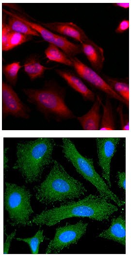
ICC/IF analysis of MAPK3 in HeLa cells line, stained with Hoechst 3342 (Blue) for nucleus staining and monoclonal anti-human MAPK3 antibody (1:500) with goat anti-mouse IgG-Texas Red conjugate (Red).
ICC/IF analysis of MAPK3 in HeLa cells line, stained with DAPI (Blue) for nucleus staining and monoclonal anti-human MAPK3 antibody (1:100) with goat anti-mouse IgG-Alexa fluor 488 conjugate (Green).
|
|
|
|
The Recombinant proteins (50ng) were resolved by SDS-PAGE, transferred to PVDF membrane and probed with anti-human MAPK3 antibody (1:500). Proteins were visualized using a goat anti-mouse secondary antibody conjugated to HRP and an ECL detection system.
Lane 1.: MAPK1 recombinant protein
Lane 2.: MAPK3 recombinant protein
The Cell lysates (40ug) were resolved by SDS-PAGE, transferred to PVDF membrane and probed with anti-human MAPK3 antibody (1:500). Proteins were visualized using a goat anti-mouse secondary antibody conjugated to HRP and an ECL detection system.
Lane 1.: HepG2 cell lysate
Lane 2.: HeLa cell lysate
|
|
|
| メーカー |
品番 |
包装 |
|
ATG
|
ATGA0191
|
50 UL
[1mg/ml]
|
※表示価格について
| 当社在庫 |
なし
|
| 納期目安 |
1週間程度
|
| 保存温度 |
-70℃
|
|

