
| 別品名 |
Interleukin-6 cytokine, IL-6, B-cell stimulatory factor 2, BSF-2, Interferon beta-2, IFN-beta-2, Hybridoma growth factor, CTL differentiation factor, CDF, Interleukin HP-1
|
| 種由来 |
Mouse
|
| 標識物 |
Unlabeled
|
| 精製度 |
Ig fraction - Ion Exchange /Gel Filtration
|
| 適用 |
Western Blot
Immunohistochemistry
|
| 免疫動物 |
Rabbit
|
| 抗体クラス |
IgG
|
| 交差種 |
Mouse
|
| GENE ID |
16193
|
| Accession No.(Gene/Protein) |
NP_112445, P08505
|
| Gene Symbol |
Il-6
|
| 形状 |
凍結乾燥品
|
| 参考文献 |
Blankenstein T., Qin Z., Li W., Diamantstein T. (1990) DNA rearrangement and constitutive expression of the interleukin 6 gene in a mouse plasmacytoma. J. Exp. Med. 171:965-970 [PubMed: 2106569] [Abstract]
|
| [注意事項] |
濃度はロットによって異なる可能性があります。メーカーDS及びCoAからご確認ください。
|
|
※サムネイル画像をクリックすると拡大画像が表示されます。
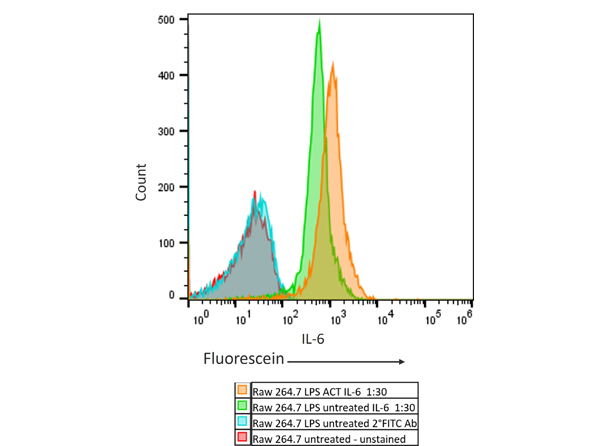
Flow Cytometry Results of Rabbit Anti Mouse IL6 Antibody in mouse Raw 264.7 cell line. The orange histogram represents the Raw 264.7 murine cells that were activated with 100ng/mL LPS for 24 hours. The green histogram are untreated Raw 264.7 cells. These two populations were intracellularly stained for 30 minutes at 4C in 1x BD Perm/WashTM buffer. The primary stain was a 1:30 dilution of the Anti Mouse IL 6 (RABBIT) Polyclonal Antibody (p/n 210 401 310, Lot#45055 [stock conc 1mg/mL]) and the secondary stain was the Anti RABBIT IgG (H&L) (GOAT) Antibody Fluorescein Conjugated (p/n 611 1202 [1:400 dilution of 2mg/mL]). The secondary stain was for 30 minutes at 4C and was kept protected from light. The blue histogram is the Raw 264.7 murine cells that were untreated and only stained with the secondary antibody. The red histogram is the untreated Raw 264.7 murine cells that were not stained. Prior to staining, the cells for all conditions were permeabilized with BD Fixation/Permeabilization TM solution for 20 minutes at 4C. All washes and stains were performed in the BD 1x Perm/WashTM buffer.
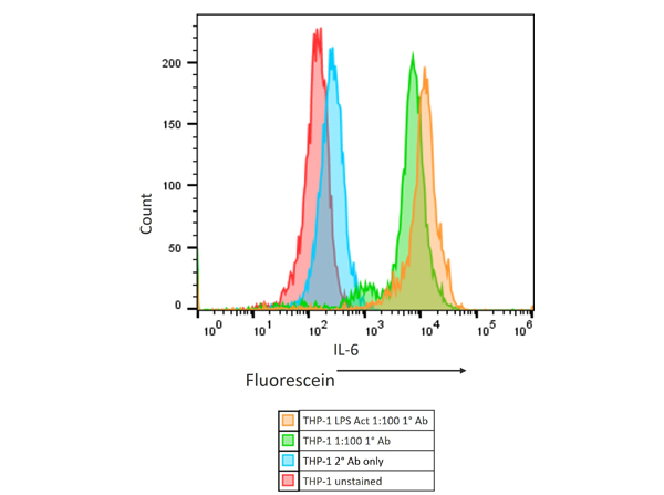
Flow Cytometry Results of Rabbit Anti-Mouse IL6 Antibody in human THP-1 cell line. The orange histogram represents the THP-1 cells that were activated with 100ng/mL LPS for 24 hours. The green histogram are untreated THP-1 cells. These two populations were intracellularly stained for 30 minutes at 4C in 1× BD Perm/WashTM buffer. The primary stain was a 1:100 dilution of the Anti-Mouse IL-6 (RABBIT) Polyclonal Antibody (p/n 210-401-310, Lot# 45055 [stock conc 1mg/mL]) and the secondary stain was the Anti-RABBIT IgG (H&L) (GOAT) Antibody Fluorescein Conjugated (p/n 611-1202 [1:400 dilution of 2mg/mL]). The secondary stain was for 30 minutes at 4C and was kept protected from light. The blue histogram is the THP-1 cells that were untreated and only stained with the secondary antibody. The red histogram is the untreated THP-1 cells that were not stained. Prior to staining, the cells for all conditions were permeabilized with BD Fixation/Permeabilization TM solution for 20 minutes at 4C. All washes and stains were performed in the BD 1× Perm/WashTM buffer.
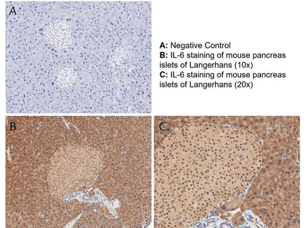
Immunohistochemistry with anti-IL-6 antibody showing positivity of islets of Langerhans (brown staining) and cytoplasmic staining in mouse pancreas at 10x and 20x (B & C). Staining was performed on Leica Bond system using the standard protocol. Formalin fixed/paraffin embedded tissue sections were subjected to antigen retrieval with E1 (Leica Microsystems) retrieval solution for 20 min and then incubated with rabbit anti-mouse IL-6 antibody at 1:50 dilution for 60 minutes. Biotinylated Anti-rabbit secondary antibody was used at 1:200 dilution to detect primary antibody. The reaction was developed using streptavidin-HRP conjugated compact polymer system and visualized with chromogen substrate, 3’3-diamino-benzidine substrate (DAB). The sections were then counterstained with hematoxylin to detect cell nuclei.
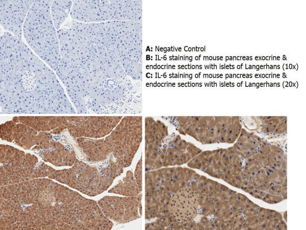
Immunohistochemistry with anti-IL-6 antibody showing cytoplasmic IL-6 staining in mouse pancreas exocrine and endocrine sections with islets of Langerhans at 10x and 20x (B & C). Staining was performed on Leica Bond system using the standard protocol. Formalin fixed/paraffin embedded tissue sections were subjected to antigen retrieval with E1 (Leica Microsystems) retrieval solution for 20 min and then incubated with rabbit anti-mouse IL-6 antibody at 1:50 dilution for 60 minutes. Biotinylated Anti-rabbit secondary antibody was used at 1:200 dilution to detect primary antibody. The reaction was developed using streptavidin-HRP conjugated compact polymer system and visualized with chromogen substrate, 3’3-diamino-benzidine substrate (DAB). The sections were then counterstained with hematoxylin to detect cell nuclei.
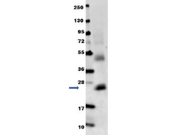
Anti-mouse IL-6 antibody in western blot shows detection of recombinant mouse IL-6 raised in E.coli. Recombinant truncated protein (0.1 ug, 21.7 kDa) was loaded on to an SDS-PAGE gel, and after separation, transferred to nitrocellulose. The membrane was blocked with 1% BSA in TBST for 30 min at RT, followed by incubation with Rockland's Anti-Mouse IL-6 antibody diluted 1:1,000 in 1% BSA in TBST overnight at 4C. After washes, the blot was reacted with secondary antibody DylightTM 649 Conjugated Anti-Rabbit IgG (H&L) (Goat) Antibody (p/n 611-143-122) diluted 1:20,000 in blocking buffer (p/n MB-070) for 30 min at RT. Data was collected using Bio-Rad VersaDocR 4000 MP imaging system.
|
|
|
|
Flow Cytometry Results of Rabbit Anti Mouse IL6 Antibody in mouse Raw 264.7 cell line. The orange histogram represents the Raw 264.7 murine cells that were activated with 100ng/mL LPS for 24 hours. The green histogram are untreated Raw 264.7 cells. These two populations were intracellularly stained for 30 minutes at 4C in 1x BD Perm/WashTM buffer. The primary stain was a 1:30 dilution of the Anti Mouse IL 6 (RABBIT) Polyclonal Antibody (p/n 210 401 310, Lot#45055 [stock conc 1mg/mL]) and the secondary stain was the Anti RABBIT IgG (H&L) (GOAT) Antibody Fluorescein Conjugated (p/n 611 1202 [1:400 dilution of 2mg/mL]). The secondary stain was for 30 minutes at 4C and was kept protected from light. The blue histogram is the Raw 264.7 murine cells that were untreated and only stained with the secondary antibody. The red histogram is the untreated Raw 264.7 murine cells that were not stained. Prior to staining, the cells for all conditions were permeabilized with BD Fixation/Permeabilization TM solution for 20 minutes at 4C. All washes and stains were performed in the BD 1x Perm/WashTM buffer.
|
|
|
| メーカー |
品番 |
包装 |
|
RKL
|
210-401-310
|
100 UG
|
※表示価格について
| 当社在庫 |
なし
|
| 納期目安 |
約10日
|
| 保存温度 |
4℃
|
|

