
| 別品名 |
Interleukin-9, IL-9, IL9, T cell growth factor 3, T cell growth factor p40, TCGF 3, Mast cell growth factor, MCGF, Megakaryoblast growth factor, p40 cytokine
|
| 種由来 |
Human
|
| 標識物 |
Unlabeled
|
| 精製度 |
Ig fraction - Ion Exchange /Gel Filtration
|
| 適用 |
Western Blot
|
| 免疫動物 |
Rabbit
|
| 抗体クラス |
IgG
|
| 交差種 |
Human
|
| GENE ID |
3578
|
| Accession No.(Gene/Protein) |
NP_000581, P15248
|
| Gene Symbol |
IL9
|
| 形状 |
凍結乾燥品
|
| 参考文献 |
Zhu,W., Liu,N., Zhao,Y., Jia,H., Cui,B. and Ning,G. (2010) Association analysis of polymorphisms in IL-3, IL-4, IL-5, IL-9 and IL-13 with Graves' disease. J. Endocrinol. Invest. (2010) In press Ciprandi,G. (2010) Serum interleukin 9 in allergic rhinitis. Ann. Allergy Asthma Immunol. 104 (2), 180-181. Osterfeld,H., Ahrens,R., Strait,R., Finkelman,F.D., Renauld,J.C. and Hogan,S.P. ((2010) Differential roles for the IL-9/IL-9 receptor alpha-chain pathway in systemic and oral antigen-induced anaphylaxis. J. Allergy Clin. Immunol. 125 (2), 469-476.
|
| [注意事項] |
濃度はロットによって異なる可能性があります。メーカーDS及びCoAからご確認ください。
|
|
※サムネイル画像をクリックすると拡大画像が表示されます。
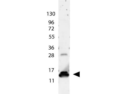
Rockland's anti-Human IL-9 antibody shows detection of a band ~15 kDa in size corresponding to recombinant human IL-9. The identity of the faint higher molecular weight band may represent a homodimer. Molecular weight markers are also shown (left). After transfer, the membrane was blocked overnight with 3% BSA in TBS followed by reaction with primary antibody at a 1:1,000 dilution. Detection occurred using peroxidase conjugated anti-Rabbit IgG (p/n 611-103-122) secondary antibody diluted 1:40,000 in blocking buffer (p/n MB-070) for 30 min at RT followed by reaction with FemtoMax? chemiluminescent substrate. Image was captured using VersaDoc? MP 4000 imaging system (Bio-Rad).
|
|
|
|
Rockland's anti-Human IL-9 antibody shows detection of a band ~15 kDa in size corresponding to recombinant human IL-9. The identity of the faint higher molecular weight band may represent a homodimer. Molecular weight markers are also shown (left). After transfer, the membrane was blocked overnight with 3% BSA in TBS followed by reaction with primary antibody at a 1:1,000 dilution. Detection occurred using peroxidase conjugated anti-Rabbit IgG (p/n 611-103-122) secondary antibody diluted 1:40,000 in blocking buffer (p/n MB-070) for 30 min at RT followed by reaction with FemtoMax? chemiluminescent substrate. Image was captured using VersaDoc? MP 4000 imaging system (Bio-Rad).
|
|
|
| メーカー |
品番 |
包装 |
|
RKL
|
209-401-B96
|
100 UG
|
※表示価格について
| 当社在庫 |
なし
|
| 納期目安 |
約10日
|
| 保存温度 |
4℃
|
|

