|
※サムネイル画像をクリックすると拡大画像が表示されます。
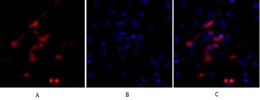
Immunofluorescence analysis of human-liver tissue. 1,p53 Polyclonal Antibody(red) was diluted at 1:200(4°C,overnight). 2, Cy3 labled Secondary antibody was diluted at 1:300(room temperature, 50min).3, Picture B: DAPI(blue) 10min. Picture A:Target. Picture B: DAPI. Picture C: merge of A+B
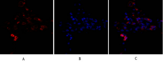
Immunofluorescence analysis of human-lung tissue. 1,p53 Polyclonal Antibody(red) was diluted at 1:200(4°C,overnight). 2, Cy3 labled Secondary antibody was diluted at 1:300(room temperature, 50min).3, Picture B: DAPI(blue) 10min. Picture A:Target. Picture B: DAPI. Picture C: merge of A+B
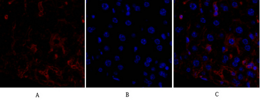
Immunofluorescence analysis of mouse-liver tissue. 1,p53 Polyclonal Antibody(red) was diluted at 1:200(4°C,overnight). 2, Cy3 labled Secondary antibody was diluted at 1:300(room temperature, 50min).3, Picture B: DAPI(blue) 10min. Picture A:Target. Picture B: DAPI. Picture C: merge of A+B
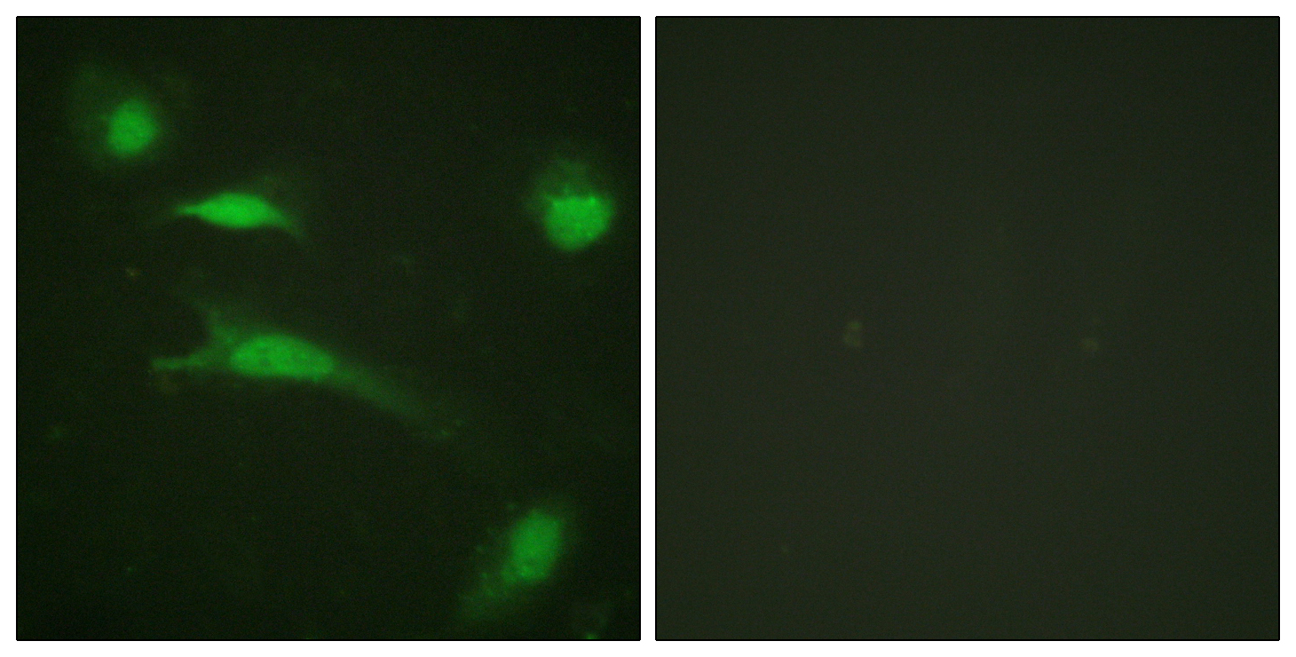
Immunofluorescence analysis of HeLa cells, using p53 Antibody. The picture on the right is blocked with the synthesized peptide.
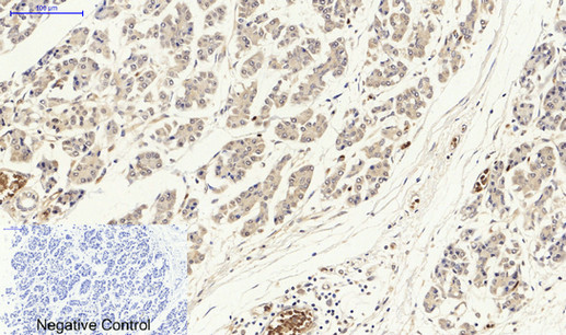
Immunohistochemical analysis of paraffin-embedded Human-lung-cancer tissue. 1,p53 Polyclonal Antibody was diluted at 1:200(4°C,overnight). 2, Sodium citrate pH 6.0 was used for antibody retrieval(>98°C,20min). 3,Secondary antibody was diluted at 1:200(room tempeRature, 30min). Negative control was used by secondary antibody only.
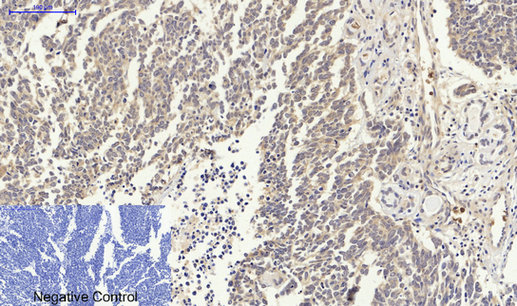
Immunohistochemical analysis of paraffin-embedded Human-stomach-cancer tissue. 1,p53 Polyclonal Antibody was diluted at 1:200(4°C,overnight). 2, Sodium citrate pH 6.0 was used for antibody retrieval(>98°C,20min). 3,Secondary antibody was diluted at 1:200(room tempeRature, 30min). Negative control was used by secondary antibody only.
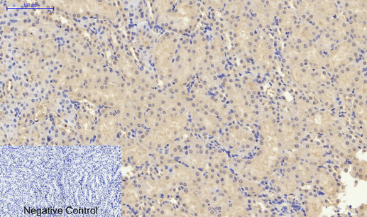
Immunohistochemical analysis of paraffin-embedded Mouse-kidney tissue. 1,p53 Polyclonal Antibody was diluted at 1:200(4°C,overnight). 2, Sodium citrate pH 6.0 was used for antibody retrieval(>98°C,20min). 3,Secondary antibody was diluted at 1:200(room tempeRature, 30min). Negative control was used by secondary antibody only.
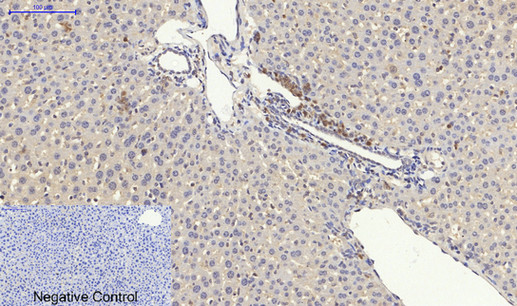
Immunohistochemical analysis of paraffin-embedded Mouse-liver tissue. 1,p53 Polyclonal Antibody was diluted at 1:200(4°C,overnight). 2, Sodium citrate pH 6.0 was used for antibody retrieval(>98°C,20min). 3,Secondary antibody was diluted at 1:200(room tempeRature, 30min). Negative control was used by secondary antibody only.
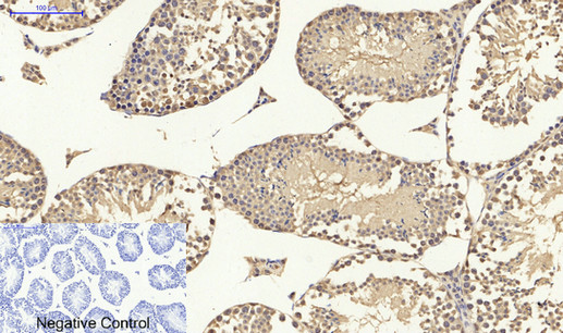
Immunohistochemical analysis of paraffin-embedded Mouse-testis tissue. 1,p53 Polyclonal Antibody was diluted at 1:200(4°C,overnight). 2, Sodium citrate pH 6.0 was used for antibody retrieval(>98°C,20min). 3,Secondary antibody was diluted at 1:200(room tempeRature, 30min). Negative control was used by secondary antibody only.
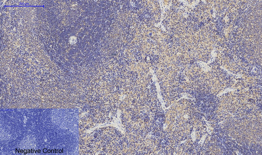
Immunohistochemical analysis of paraffin-embedded Rat-spleen tissue. 1,p53 Polyclonal Antibody was diluted at 1:200(4°C,overnight). 2, Sodium citrate pH 6.0 was used for antibody retrieval(>98°C,20min). 3,Secondary antibody was diluted at 1:200(room tempeRature, 30min). Negative control was used by secondary antibody only.
|
|
|
|
Immunofluorescence analysis of human-liver tissue. 1,p53 Polyclonal Antibody(red) was diluted at 1:200(4°C,overnight). 2, Cy3 labled Secondary antibody was diluted at 1:300(room temperature, 50min).3, Picture B: DAPI(blue) 10min. Picture A:Target. Picture B: DAPI. Picture C: merge of A+B
|
|
| 別品名 |
Antigen NY-CO-13; Cellular tumor antigen p53; Phosphoprotein p53; TP53; tumor protein p53; Tumor suppressor p53
|
| 種由来 |
Human
|
| 交差種 |
Human
Mouse
Rat
Monkey
|
| 適用 |
Western Blot
IHC paraffin embedding section
Enzyme Linked Immunosorbent Assay
Immuno Fluorescence
Immunocytochemistry (cell)
|
| 免疫動物 |
Rabbit
|
| 抗体クラス |
IgG
|
| 抗原部位 |
a.a.10-59
|
| 標識物 |
Unlabeled
|
| 精製度 |
Affinity Purified
|
| GENE ID |
7157
|
| Accession No.(Gene/Protein) |
P04637
|
| Gene Symbol |
TP53
|
| 分子量 |
43 kDa
|
|
| メーカー |
品番 |
包装 |
|
ASY
|
B0529
|
100 UL
[1 mg/ml]
|
※表示価格について
| 当社在庫 |
なし
|
| 納期目安 |
2週間程度
|
| 保存温度 |
-20℃
|
|
