|
※サムネイル画像をクリックすると拡大画像が表示されます。
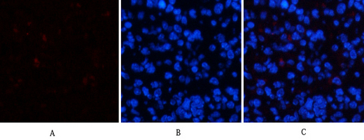
Immunofluorescence analysis of mouse-spleen tissue. 1,ERK 1/2 (phospho Tyr204) Polyclonal Antibody(red) was diluted at 1:200(4°C,overnight). 2, Cy3 labled Secondary antibody was diluted at 1:300(room temperature, 50min).3, Picture B: DAPI(blue) 10min. Picture A:Target. Picture B: DAPI. Picture C: merge of A+B
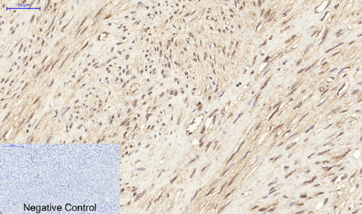
Immunohistochemical analysis of paraffin-embedded Human-uterus tissue. 1,ERK 1/2 (phospho Tyr204) Polyclonal Antibody was diluted at 1:200(4°C,overnight). 2, Sodium citrate pH 6.0 was used for antibody retrieval(>98°C,20min). 3,Secondary antibody was diluted at 1:200(room tempeRature, 30min). Negative control was used by secondary antibody only.
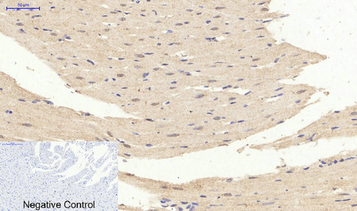
Immunohistochemical analysis of paraffin-embedded Rat-heart tissue. 1,ERK 1/2 (phospho Tyr204) Polyclonal Antibody was diluted at 1:200(4°C,overnight). 2, Sodium citrate pH 6.0 was used for antibody retrieval(>98°C,20min). 3,Secondary antibody was diluted at 1:200(room tempeRature, 30min). Negative control was used by secondary antibody only.
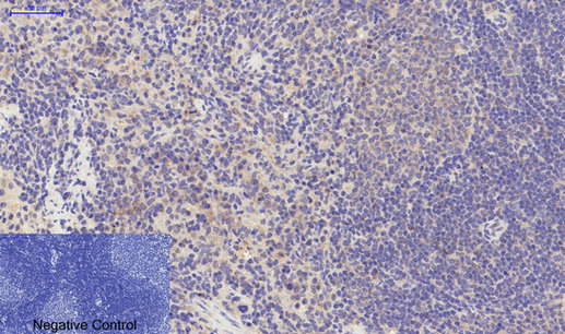
Immunohistochemical analysis of paraffin-embedded Rat-spleen tissue. 1,ERK 1/2 (phospho Tyr204) Polyclonal Antibody was diluted at 1:200(4°C,overnight). 2, Sodium citrate pH 6.0 was used for antibody retrieval(>98°C,20min). 3,Secondary antibody was diluted at 1:200(room tempeRature, 30min). Negative control was used by secondary antibody only.
Immunohistochemical analysis of paraffin-embedded Human brain. Antibody was diluted at 1:100(4° overnight). High-pressure and temperature Tris-EDTA,pH8.0 was used for antigen retrieval. Negetive contrl (right) obtaned from antibody was pre-absorbed by immunogen peptide.

Immunohistochemistry analysis of paraffin-embedded human breast carcinoma, using p44/42 MAP Kinase (Phospho-Tyr204) Antibody. The picture on the right is blocked with the phospho peptide.

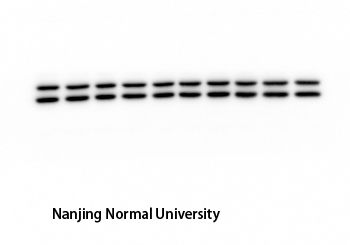
Western Blot analysis of HeLa cells using Phospho-ERK 1/2 (Y204) Polyclonal Antibody diluted at 1:2000

Western Blot analysis of various cells using Phospho-ERK 1/2 (Y204) Polyclonal Antibody diluted at 1:2000

Western blot analysis of lysates from HeLa cells treated with EGF 200ng/ml 30', using p44/42 MAP Kinase (Phospho-Tyr204) Antibody. The lane on the right is blocked with the phospho peptide.

The picture was kindly provided by our customer
|
|
|
|
Immunofluorescence analysis of mouse-spleen tissue. 1,ERK 1/2 (phospho Tyr204) Polyclonal Antibody(red) was diluted at 1:200(4°C,overnight). 2, Cy3 labled Secondary antibody was diluted at 1:300(room temperature, 50min).3, Picture B: DAPI(blue) 10min. Picture A:Target. Picture B: DAPI. Picture C: merge of A+B
|
|
| 別品名 |
ERK-1; ERK1; ERT2; Extracellular signal- regulated kinase 1; Insulin-stimulated MAP2 kinase; kinase ERK1; MAP kinase 1; MAPK 1; MAPK3; Microtubule- associated protein-2 kinase; Mitogen-activated protein kinase 3; MK03; MNK1; p44-ERK1; P44-ERK1; p44-MAPK;
|
| 種由来 |
Human
|
| 交差種 |
Human
Mouse
Rat
|
| 適用 |
Western Blot
IHC paraffin embedding section
Enzyme Linked Immunosorbent Assay
Immuno Fluorescence
Immunocytochemistry (cell)
|
| 免疫動物 |
Rabbit
|
| 抗体クラス |
IgG
|
| 抗原部位 |
a.a.170-219
|
| 標識物 |
Unlabeled
|
| 精製度 |
Affinity Purified
|
| 翻訳後修飾 |
リン酸化
|
| GENE ID |
5595/5594
|
| Accession No.(Gene/Protein) |
P27361, P28482
|
| Gene Symbol |
MAPK3
|
| 分子量 |
43 kDa
|
|
| メーカー |
品番 |
包装 |
|
ASY
|
A7074
|
100 UL
[1 mg/ml]
|
※表示価格について
| 当社在庫 |
なし
|
| 納期目安 |
2週間程度
|
| 保存温度 |
-20℃
|
|
※当社では商品情報の適切な管理に努めておりますが、表示される法規制情報は最新でない可能性があります。
また法規制情報の表示が無いものは、必ずしも法規制に非該当であることを示すものではありません。
商品のお届け前に最新の製品法規制情報をお求めの際はこちらへお問い合わせください。
|
※当社取り扱いの試薬・機器製品および受託サービス・創薬支援サービス(納品物、解析データ等)は、研究用としてのみ販売しております。
人や動物の医療用・臨床診断用・食品用としては、使用しないように、十分ご注意ください。
法規制欄に体外診断用医薬品と記載のものは除きます。
|
|
※リンク先での文献等のダウンロードに際しましては、掲載元の規約遵守をお願いします。
|
