|
※サムネイル画像をクリックすると拡大画像が表示されます。
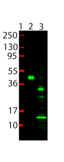
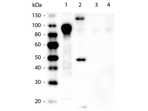
Western Blot showing detection of 6x HIS Epitope Tag. Lane 1: Molecular Weight Marker. Lane 2: Recombinant 6xHIS-SUMO-GFP. Lane 3: Nag1. Blocking with 5% Blotto (p/n B501-0500) 30 min at 20°C. Detection: Anti-6x His Epitope Tag (MOUSE) Monoclonal Antibody DyLight? 800 Conjugated (p/n 200-345-382) secondary antibody was used at 1:5000 in Blocking Buffer for Fluorescent Western Blotting (p/n MB-070) and imaged on the LiCor Odyssey imaging system.
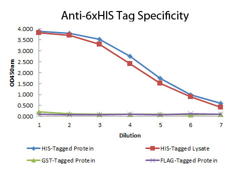
ELISA of Mouse anti-6xHIS Tag Antibody. Antigen: HIS-tagged purified protein and E. coli cell lysates expressing HIS-Tagged construct, GST- and RON-tagged purified proteins. Coating amount: 0.15ug per well. Primary antibody: 6xHIS Tag antibody at 100ug/mL. Dilution series: 2-fold. Mid-point concentration: 200ng/mL. Secondary antibody: Peroxidase mouse secondary antibody at 1:10,000. Substrate: TMB (p/n TMBE-1000).
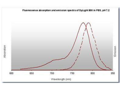
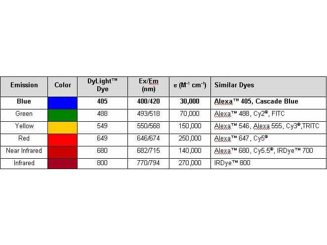
Properties of DyLight? Conjugates.
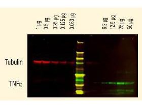
DyLight? dyes can be used for two-color Western Blot detection with low background and high signal. Anti-tubulin was detected using a DyLight? 680 conjugate. Anti-TNFα was detected using a DyLight? 800 conjugate. The image was captured using the OdysseyR Infrared Imaging System developed by LI-COR.
Western Blot of Mouse anti-6xHIS Tag Antibody. Lane 1: 100ng Purified histidine-tagged recombinant protein. Lane 2: 200ng E. coli cell lysate containing histidine-tagged expression construct. Lane 3: 100ng Purified GST-tagged recombinant protein. Lane 4: 100ng Purified FLAG-tagged recombinant protein. Primary antibody: Mouse anti-6xHIS Tag antibody at 1:5,000 overnight at 4°C. Secondary antibody: Peroxidase mouse secondary antibody at 1:20,000 for 30 min at RT. Block: 5% BLOTTO for 1 hr at RT.
|
|
|
|
Western Blot showing detection of 6x HIS Epitope Tag. Lane 1: Molecular Weight Marker. Lane 2: Recombinant 6xHIS-SUMO-GFP. Lane 3: Nag1. Blocking with 5% Blotto (p/n B501-0500) 30 min at 20°C. Detection: Anti-6x His Epitope Tag (MOUSE) Monoclonal Antibody DyLight? 800 Conjugated (p/n 200-345-382) secondary antibody was used at 1:5000 in Blocking Buffer for Fluorescent Western Blotting (p/n MB-070) and imaged on the LiCor Odyssey imaging system.
|
|
| 別品名 |
mouse anti-6X His Tag DyLightTM 800 conjugated Antibody, DyLightTM 800 conjugated mouse anti-6X His Tag Antibody, anti-HIS, HIS Antibody, 6X His Tag Antibody, HHHHHH epitope tag antibody
|
| 適用 |
Western Blot
Enzyme Linked Immunosorbent Assay
Dot Blot
|
| 免疫動物 |
Mouse
|
| クローン |
33D10.D2.G8
|
| 抗体クラス |
IgG1κ
|
| 標識物 |
DyLightTM 800
|
| 精製度 |
Ig fraction - Protein A
|
| Tag情報 |
6X HIS
|
| 参考文献 |
[Pub Med ID]35487399
|
| [注意事項] |
濃度はロットによって異なる可能性があります。メーカーDS及びCoAからご確認ください。
|
|
| メーカー |
品番 |
包装 |
|
RKL
|
200-345-382
|
100 UG
|
※表示価格について
| 当社在庫 |
なし
|
| 納期目安 |
約10日
|
| 法規制 |
毒
|
| 保存温度 |
4℃
|
|
※当社では商品情報の適切な管理に努めておりますが、表示される法規制情報は最新でない可能性があります。
また法規制情報の表示が無いものは、必ずしも法規制に非該当であることを示すものではありません。
商品のお届け前に最新の製品法規制情報をお求めの際はこちらへお問い合わせください。
|
※当社取り扱いの試薬・機器製品および受託サービス・創薬支援サービス(納品物、解析データ等)は、研究用としてのみ販売しております。
人や動物の医療用・臨床診断用・食品用としては、使用しないように、十分ご注意ください。
法規制欄に体外診断用医薬品と記載のものは除きます。
|
|
※リンク先での文献等のダウンロードに際しましては、掲載元の規約遵守をお願いします。
|
|
※CAS Registry Numbers have not been verified by CAS and may be inaccurate.
|