|
※サムネイル画像をクリックすると拡大画像が表示されます。
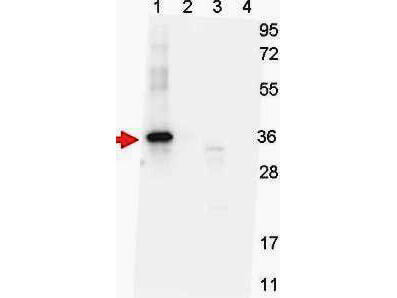
Western blot shows detection of recombinant NAG-1 protein present in Pichia pastoris whole cell lysates: lane 1 - yeast cell lysate expressing NAG-1 H variant with SUMO expression tag at 36 kDa; lane 2 - yeast cell lysate expressing NAG-1 D variant with SUMO expression tag at 36 kDa; lane 3 - yeast cell lysate expressing NAG-1 H variant; and lane 4 - yeast cell lysate expressing NAG-1 D variant. All lysates were run under reducing conditions. Primary antibody was used at a 1:1,000 dilution in TBS containing 1% BSA and 0.2% Tween, and reacted overnight at 4°C. For detection, a 1:40,000 dilution of peroxidase conjugated Gt-a-Mouse IgG secondary antibody (610-103-121) was used in Blocking Buffer for Fluorescent Western Blotting (MB-070) for 30 min at room temperature. Molecular weight estimation was made by comparison to prestained MW markers. Image was captured using the BioRad Versadoc? 4000MP Imaging System.
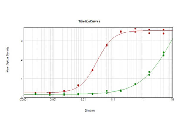
ELISA Results of Mouse Anti-NAG-1 H variant Antibody. Each well was coated in duplicate with 0.1μg of conjugate [H-variant Red line, D-variant Green line]. The working dilution is 1:35,000. The starting dilution of antibody was 5μg/ml and the X-axis represents the Log10 of a 3-fold dilution. This titration is a 4-parameter curve fit where the IC50 is defined as the titer of the antibody. Assay performed using HRP Conjugate Stabilizer (p/n MB-076), Rabbit Anti-Mouse IgG HRP conjugated (p/n 610-403-C46) and TMB substrate (p/n TMBE-1000).
|
|
|
|
Western blot shows detection of recombinant NAG-1 protein present in Pichia pastoris whole cell lysates: lane 1 - yeast cell lysate expressing NAG-1 H variant with SUMO expression tag at 36 kDa; lane 2 - yeast cell lysate expressing NAG-1 D variant with SUMO expression tag at 36 kDa; lane 3 - yeast cell lysate expressing NAG-1 H variant; and lane 4 - yeast cell lysate expressing NAG-1 D variant. All lysates were run under reducing conditions. Primary antibody was used at a 1:1,000 dilution in TBS containing 1% BSA and 0.2% Tween, and reacted overnight at 4°C. For detection, a 1:40,000 dilution of peroxidase conjugated Gt-a-Mouse IgG secondary antibody (610-103-121) was used in Blocking Buffer for Fluorescent Western Blotting (MB-070) for 30 min at room temperature. Molecular weight estimation was made by comparison to prestained MW markers. Image was captured using the BioRad Versadoc? 4000MP Imaging System.
|
|
| 別品名 |
mouse anti-NAG1 Antibody, NAG-1, GDF15, MIC-1, nonsteroidal anti-inflammatory drug-activated gene, NSAID-activated gene 1 protein, growth differentiation factor 15, macrophage inhibitory compound 1, prostate-derived factor
|
| 交差種 |
Human
|
| 適用 |
Western Blot
Enzyme Linked Immunosorbent Assay
|
| 免疫動物 |
Mouse
|
| クローン |
7C12.B3.F2
|
| 抗体クラス |
IgG2bκ
|
| 抗原部位 |
N-terminus
|
| 標識物 |
Unlabeled
|
| 精製度 |
Ig fraction - Protein A
|
| GENE ID |
9518
|
| Accession No.(Gene/Protein) |
Q99988.3, Q99988
|
| Gene Symbol |
GDF15
|
| 参考文献 |
Baek, S.J., Eling, T.E. (2006) Changes in gene expression contribute to cancer prevention by COX inhibitors. Prog Lipid Res. 45(1):1-16. Lindmark, F., Zheng, S.L., Wiklund, F., Bensen, J., Balter, K.A., Chang, B., Hedelin, M., Clark, J., Stattin, P., Meyers, D.A., Adami, H-O., Isaacs, W., Gronberg, H. and Xu, J. (2004) H6D Polymorphism in Macrophage-Inhibitory Cytokine-1 Gene Associated With Prostate Cancer J Natl Cancer Inst. 96(16): 1248-1254.
|
| [注意事項] |
濃度はロットによって異なる可能性があります。メーカーDS及びCoAからご確認ください。
|
|
| メーカー |
品番 |
包装 |
|
RKL
|
200-301-B08
|
100 UG
|
※表示価格について
| 当社在庫 |
なし
|
| 納期目安 |
約10日
|
| 保存温度 |
-20℃
|
|
※当社では商品情報の適切な管理に努めておりますが、表示される法規制情報は最新でない可能性があります。
また法規制情報の表示が無いものは、必ずしも法規制に非該当であることを示すものではありません。
商品のお届け前に最新の製品法規制情報をお求めの際はこちらへお問い合わせください。
|
※当社取り扱いの試薬・機器製品および受託サービス・創薬支援サービス(納品物、解析データ等)は、研究用としてのみ販売しております。
人や動物の医療用・臨床診断用・食品用としては、使用しないように、十分ご注意ください。
法規制欄に体外診断用医薬品と記載のものは除きます。
|
|
※リンク先での文献等のダウンロードに際しましては、掲載元の規約遵守をお願いします。
|
|
※CAS Registry Numbers have not been verified by CAS and may be inaccurate.
|
