|
※サムネイル画像をクリックすると拡大画像が表示されます。
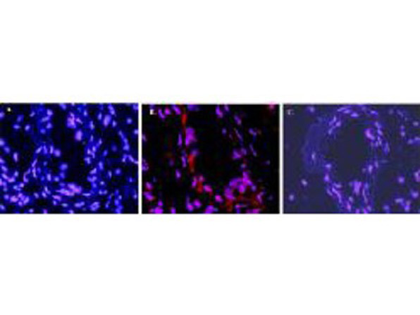
Immunofluorescence microscopy after staining of mouse carotid artery tissue with anti-Mouse IL-1s antiserum (p/n 110-401-319, less purified form of 210-401-319) diluted 1:50. Tissue sections were prepared after cryofixation. Reaction occurred at room temperature for 60' followed by washes and reaction with Rhodamine conjugated Gt-a-Rabbit IgG (p/n 611-100-122). Tissue was counterstained with bis-benzimide solution at 0.5 mg/ml in PBS for 15 min at room temperature. Panel A) shows no antibody staining of WT uninjured mouse carotid tissue. Panel B) shows anti-IL-1s staining of cells after surgical injury of tissue. Panel C) shows no antibody staining of injured carotid tissue from an IL-1s KO mouse.
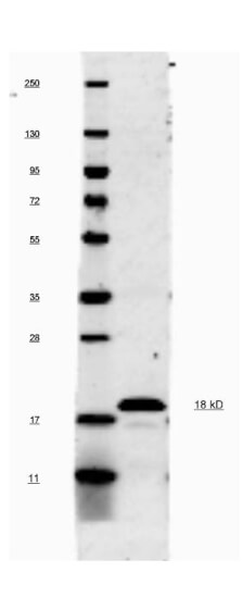
This antibody will recognize 10% of the non-denatured (native) precursor 31,000 MW mouse IL-1s containing samples but will primarily detect all of the 17,000 MW mature molecule. However, in western blot analysis, the usual procedure of heating the sample in SDS with or without reducing agents will facilitate denaturing of the 31,000 MW IL- 1s precursor molecule. Denatured IL-1s will have a 18 kDa band.
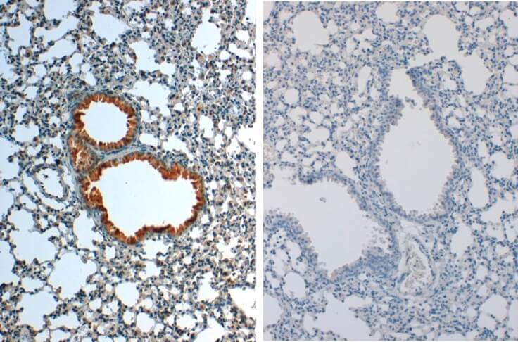
Immunohistochemistry of Rabbit anti-IL1Beta Antibody in Mouse Embryonic Kidney Tissue: Mouse Embryonic Kidney Fixation: FFPE buffered formalin 10% conc Ag Retrieval: Heat, Citrate pH 6.2. Pressure Cooker Primary antibody: 2ug/ml 1.5 hour @ room T Secondary Ab: MACH 1 HRP POLYMER 1:50 45” RT
|
|
|
|
Immunofluorescence microscopy after staining of mouse carotid artery tissue with anti-Mouse IL-1s antiserum (p/n 110-401-319, less purified form of 210-401-319) diluted 1:50. Tissue sections were prepared after cryofixation. Reaction occurred at room temperature for 60' followed by washes and reaction with Rhodamine conjugated Gt-a-Rabbit IgG (p/n 611-100-122). Tissue was counterstained with bis-benzimide solution at 0.5 mg/ml in PBS for 15 min at room temperature. Panel A) shows no antibody staining of WT uninjured mouse carotid tissue. Panel B) shows anti-IL-1s staining of cells after surgical injury of tissue. Panel C) shows no antibody staining of injured carotid tissue from an IL-1s KO mouse.
|
|
| 別品名 |
rabbit anti-IL-1 beta antibody, rabbit anti-IL-1b antibody, rabbit anti-Interleukin-1 beta antibody, IL-1β, catabolin
|
| 交差種 |
Mouse
|
| 適用 |
Western Blot
Immunohistochemistry
Immuno Fluorescence
|
| 免疫動物 |
Rabbit
|
| 標識物 |
Unlabeled
|
| 精製度 |
Ig fraction - Ion Exchange /Gel Filtration
|
| GENE ID |
16176
|
| Accession No.(Gene/Protein) |
CAA28637.1, P10749
|
| Gene Symbol |
Il1b
|
| 参考文献 |
[Pub Med ID]32211925
|
| [注意事項] |
濃度はロットによって異なる可能性があります。メーカーDS及びCoAからご確認ください。
|
|
| メーカー |
品番 |
包装 |
|
RKL
|
210-401-319
|
100 UG
|
※表示価格について
| 当社在庫 |
なし
|
| 納期目安 |
約10日
|
| 保存温度 |
4℃
|
|
※当社では商品情報の適切な管理に努めておりますが、表示される法規制情報は最新でない可能性があります。
また法規制情報の表示が無いものは、必ずしも法規制に非該当であることを示すものではありません。
商品のお届け前に最新の製品法規制情報をお求めの際はこちらへお問い合わせください。
|
※当社取り扱いの試薬・機器製品および受託サービス・創薬支援サービス(納品物、解析データ等)は、研究用としてのみ販売しております。
人や動物の医療用・臨床診断用・食品用としては、使用しないように、十分ご注意ください。
法規制欄に体外診断用医薬品と記載のものは除きます。
|
|
※リンク先での文献等のダウンロードに際しましては、掲載元の規約遵守をお願いします。
|
|
※CAS Registry Numbers have not been verified by CAS and may be inaccurate.
|
