
| 別品名 |
HCLS1 associated protein X-1, HS1BP1, HCLSBP1, HCLS1 (and PKD2) associated protein
|
| 抗原部位 |
a.a.1-279
|
| 種由来 |
Human
|
| 標識物 |
Unlabeled
|
| 精製度 |
Ig fraction - Protein G
|
| 適用 |
Western Blot
Enzyme Linked Immunosorbent Assay
Immuno Fluorescence
Immunocytochemistry (cell)
|
| 免疫動物 |
Mouse
|
| 抗体クラス |
IgG2bκ
|
| クローン |
AT3C5
|
| 交差種 |
Human
|
| Accession No.(Gene/Protein) |
NP_006109, O00165
|
| 形状 |
液状
|
| 参考文献 |
Klein C, et al., (2007) Nat Genet. 39(1):86-92
Yin H, et al., (2001) Cytokine. 15(3):122-37
|
|
※サムネイル画像をクリックすると拡大画像が表示されます。
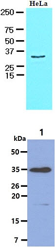
The HeLa cell lysate (30ug) were resolved by SDS-PAGE, transferred to NC membrane and probed with anti-human HAX1 antibody (1:1000). Proteins were visualized using a goat anti-mouse secondary antibody conjugated to HRP and an ECL detection system.
The Cell lysates (40ug) were resolved by SDS-PAGE, transferred to PVDF membrane and probed with anti-human HAX1 antibody (1:1000). Proteins were visualized using a goat anti-mouse secondary antibody conjugated to HRP and an ECL detection system.
Lane 1.: Raji cell lysate
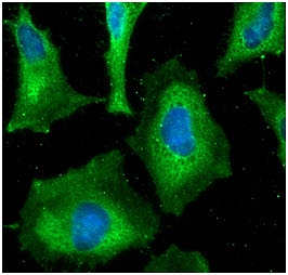
ICC/IF analysis of HAX1 in HeLa cells line, stained with DAPI (Blue) for nucleus staining and monoclonal anti-human HAX1 antibody (1:100) with goat anti-mouse IgG-Alexa fluor 488 conjugate (Green).
|
|
|
|
The HeLa cell lysate (30ug) were resolved by SDS-PAGE, transferred to NC membrane and probed with anti-human HAX1 antibody (1:1000). Proteins were visualized using a goat anti-mouse secondary antibody conjugated to HRP and an ECL detection system.
The Cell lysates (40ug) were resolved by SDS-PAGE, transferred to PVDF membrane and probed with anti-human HAX1 antibody (1:1000). Proteins were visualized using a goat anti-mouse secondary antibody conjugated to HRP and an ECL detection system.
Lane 1.: Raji cell lysate
|
|
|
| メーカー |
品番 |
包装 |
|
ATG
|
ATGA0156
|
50 UL
[1mg/ml]
|
※表示価格について
| 当社在庫 |
なし
|
| 納期目安 |
1週間程度
|
| 保存温度 |
-70℃
|
|

