

| 標識物 |
Unlabeled
|
| 精製度 |
Affinity Purified
|
| 適用 |
Western Blot
Enzyme Linked Immunosorbent Assay
Immunocytochemistry (cell)
Immunoprecipitation
|
| 免疫動物 |
Rabbit
|
| クローン |
G15-B
|
| 交差種 |
Human
Mouse
Rat
|
| 翻訳後修飾 |
リン酸化
|
| その他 |
[Uniprot ID]P28482, P63086, P63085
|
| 参考文献 |
W.R. Lee et al (2015) J Transl Med, 13:191; C.J. Tai et al (2013) Ann Diagn Pathol 17, 165-71; C.J. Tai et al (2012) Pol J Pathol 63, 93-100; E. Ranzato et al (2010) Cell Biochem Biophys 57, 9-17; J.G. Doria et al (2015) Neurobiol Dis 73, 163-73; W.R. Lee et al (2015) Mol Carcinog; L.A. Venegas et al (2016) N Biotechnol 33, 537-43; M.C. Jiang (2013) Microvesicle membrane protein and application thereof (Patent); M.C. Jiang (2013) Microvesicle protein for cancer targeting (Patent)
|
|
※サムネイル画像をクリックすると拡大画像が表示されます。
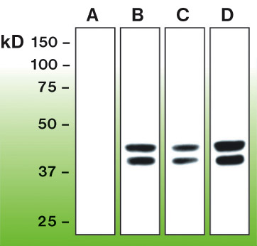
Anti-phospho-Erk 1,2 (DB 013). Western blot analysis of Erk 1,2 activation in untreated PC12 cells (A), cells treated with EGF- 100ng/ml, 5min (B), PMA-100nM, 30min (C) in serum free DMEM, and EGF-100ng/ml, 5min with 10% FBS in DMEM (D). Wells were equally loaded with 50μg of whole cell lysate proteins/well.
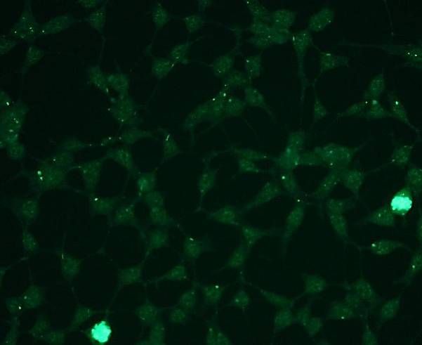
Representative pictures of phospho-Erk1,2 expression in HEK293 cells, (untreated cells) visualized with clonal rabbit anti-phospho-Erk1,2 monospecific antibody. Primary antibody dilution - 1:100.
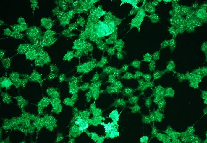
Representative pictures of phospho-Erk1,2 expression in HEK293 cells, (hydrogen peroxide treated cells) visualized with clonal rabbit anti-phospho-Erk1,2 monospecific antibody. Primary antibody dilution - 1:100.
|
|
|
|
Anti-phospho-Erk 1,2 (DB 013). Western blot analysis of Erk 1,2 activation in untreated PC12 cells (A), cells treated with EGF- 100ng/ml, 5min (B), PMA-100nM, 30min (C) in serum free DMEM, and EGF-100ng/ml, 5min with 10% FBS in DMEM (D). Wells were equally loaded with 50μg of whole cell lysate proteins/well.
|
|
|
| メーカー |
品番 |
包装 |
|
DBB
|
DB 013-0.1
|
100 UL
[4.0 mg/ml]
|
※表示価格について
| 当社在庫 |
なし
|
| 納期目安 |
約10日
|
| 保存温度 |
-20℃
|
|

