|
※サムネイル画像をクリックすると拡大画像が表示されます。
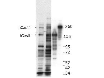
Western blot using Rockland's anti-hCASZ1 antibody. This blot shows detection of endogenous and transfected human CASZ1 protein in fresh whole cell lysate (~30 μg). Lane 1: BE2(s) cell lysate. Lane 2: BE2(N) cell lysate. Lane 3: SY5Y transfected with hCASZ5 (125kDa). Lane 4: SY5Y transfected with hCASZ11 (190kDa). Protein was resolved by SDS-PAGE and transferred onto nitrocellulose. After blocking, the membrane was probed with the primary antibody diluted to 1:1,000 for 1.5 hours at room temperature then incubated with HRP-conjugated Goat Anti-Rabbit antibody for 45 min. at room temperature. Personal communication, Carol Thiele, NCI, Bethesda, MD.
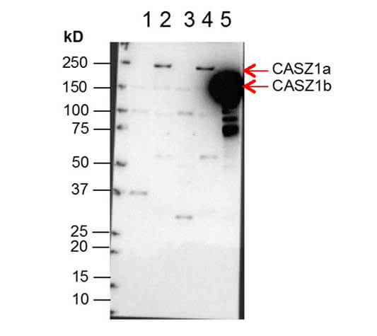
Western Blot of Anti-CASZ1 Antibody. Lane 1: NBLS Cytoplasmic (20μg). Lane 2: NBLS Nuclear (3μg). Lane 3: BE2C Cytoplasmic (30μg). Lane 4: BE2C Nuclear (7μg). Lane 5: SY5Y-CASZ1b (10μg). Block: 5% Blotto/TTBS for 1 hour. Primary: Casz1 1:10,000 for 1 hour. Secondary: Goat anti-Rabbit HRP for 1 hour. 240sec exposure. Detects nuclear endogenous CASZ1a and CASZ1b; and transiently transfected CASZ1b isoform. Personal communication and images from Carol Thiele Galetto, NCI.
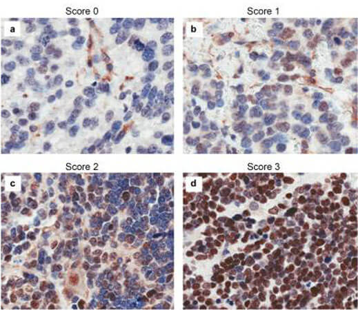
Immunohistochemistry results of Rabbit Anti-hCASZ1 Antibody. Tissue: NB patient tumor. A. Score 0- a rare positive nuclei. B. Score 1- (1-10% positive) equivocal/uninterpretable. C. Score 2- (10-50% positive) weak positive. D. Score 3- (>50% positive) strong positive. Primary Antibody: Rabbit Anti-CASZ1 stained brown. Nucleus counterstained with hematoxylin (blue). Localization: Nuclear.
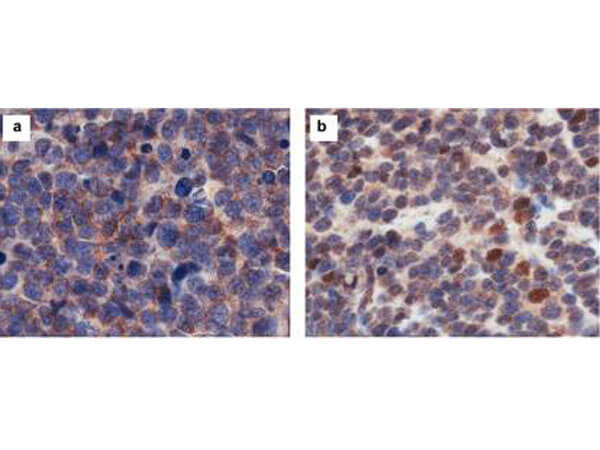
Immunohistochemistry results of Rabbit Anti-hCasz1 Antibody. Tissue: NB patient tumor. A. CASZ1 localized exclusively in the cytoplasm. B. CASZ1 localized in the cytoplasm and nucleus. Primary Antibody: Rabbit Anti-CASZ1 stained brown. Nucleus counterstained with hematoxylin (blue).
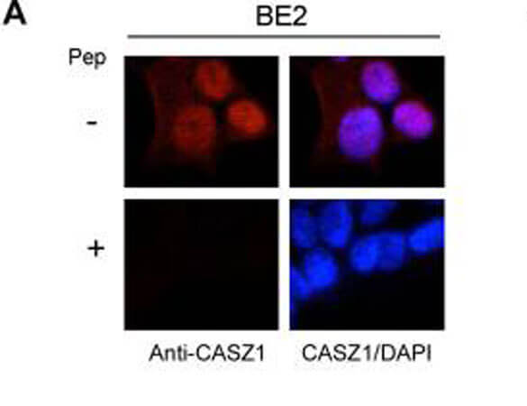
Immunofluorescence results of Endogenous CASZ1. Cells: BE2 cells. With or without Pre-Incubation of Anti-CASZ1 Antibody with CASZ1 Peptide. Staining: Rabbit Anti-CASZ1 Antibody. Chromatin counter stain: DAPI.
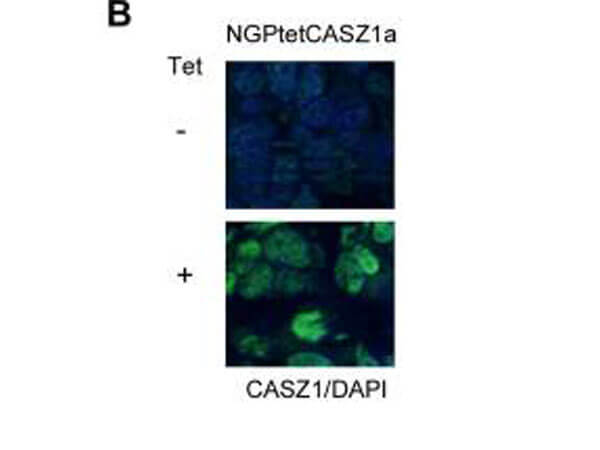
Immunofluorescence results of Rabbit Anti-CASZ1 Antibody. Tissue: Mouse Xenograft tumor of human NB cell line transfected with or without tetracycline inducible CASZ1 (NGPtetCASZ1a). Antibody: Rabbit Anti-CASZ1 Antibody. Counterstain: DAPI.
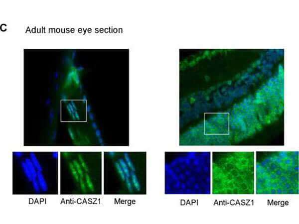
Immunofluorescence of Rabbit anti-CASZ1 Antibody. Tissue: adult murine ocular tissue. Antibody: Rabbit Anti-CASZ1 Antibody. Counterstain: DAPI. Localization: nucleus in lens epithelia but primarily localizes in the cytoplasm in photoreceptor cells.
|
|
|
|
Western blot using Rockland's anti-hCASZ1 antibody. This blot shows detection of endogenous and transfected human CASZ1 protein in fresh whole cell lysate (~30 μg). Lane 1: BE2(s) cell lysate. Lane 2: BE2(N) cell lysate. Lane 3: SY5Y transfected with hCASZ5 (125kDa). Lane 4: SY5Y transfected with hCASZ11 (190kDa). Protein was resolved by SDS-PAGE and transferred onto nitrocellulose. After blocking, the membrane was probed with the primary antibody diluted to 1:1,000 for 1.5 hours at room temperature then incubated with HRP-conjugated Goat Anti-Rabbit antibody for 45 min. at room temperature. Personal communication, Carol Thiele, NCI, Bethesda, MD.
|
|
| 別品名 |
rabbit anti-CASZ1 Antibody, CASZ1, Zinc finger protein castor homolog 1, Castor-related protein, Zinc finger protein 693, CST, SRG, ZNF693
|
| 交差種 |
Human
|
| 適用 |
Western Blot
Enzyme Linked Immunosorbent Assay
|
| 免疫動物 |
Rabbit
|
| 標識物 |
Unlabeled
|
| 精製度 |
Affinity Purified
|
| GENE ID |
54897
|
| Accession No.(Gene/Protein) |
145207289, Q86V15
|
| Gene Symbol |
CASZ1
|
| 参考文献 |
[Pub Med ID]32060262
|
| [注意事項] |
濃度はロットによって異なる可能性があります。メーカーDS及びCoAからご確認ください。
|
|
| メーカー |
品番 |
包装 |
|
RKL
|
600-401-B62S
|
25 UL
|
※表示価格について
| 当社在庫 |
なし
|
| 納期目安 |
約10日
|
| 保存温度 |
-20℃
|
|
※当社では商品情報の適切な管理に努めておりますが、表示される法規制情報は最新でない可能性があります。
また法規制情報の表示が無いものは、必ずしも法規制に非該当であることを示すものではありません。
商品のお届け前に最新の製品法規制情報をお求めの際はこちらへお問い合わせください。
|
※当社取り扱いの試薬・機器製品および受託サービス・創薬支援サービス(納品物、解析データ等)は、研究用としてのみ販売しております。
人や動物の医療用・臨床診断用・食品用としては、使用しないように、十分ご注意ください。
法規制欄に体外診断用医薬品と記載のものは除きます。
|
|
※リンク先での文献等のダウンロードに際しましては、掲載元の規約遵守をお願いします。
|
|
※CAS Registry Numbers have not been verified by CAS and may be inaccurate.
|
