|
※サムネイル画像をクリックすると拡大画像が表示されます。
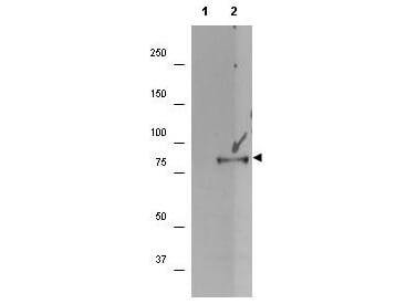
Western blot using Rockland's affinity purified anti-p90 RSK1 pS732 antibody shows detection of a band ~90 kDa in size corresponding to phosphorylated p90 RSK1 (arrowhead) in EGF stimulated (lane 2) HEK293T cell lysates prepared from cells grown in the absence of serum for 12 h. No staining is observed in similarly prepared lysates derived from unstimulated (control) cells (lane 1). After transfer, the membrane was blocked overnight followed by reaction with the primary antibody at a 1:1,000 dilution. Detection occurred using a peroxidase conjugated secondary antibody and ECL. Personal Communication. Kuldeep Patel, Loyola University Medical Center, Maywood, IL.
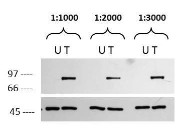
Western blot using Rockland's affinity purified anti-p90 RSK1 pS732 antibody. Lane 1-2: HEK293T (U) untreated or (T) treated with EGF. Lane 3-4: HEK293T (U) untreated or (T) treated with EGF. Lane 5-6: HEK293T (U) untreated or (T) treated with EGF. Load: 15μg per lane. Actin used as a loading control. Blocking: 5% milk. Primary Antibody: Anti-RSK1-pS732 1:1000, 1:2000, or 1:3000 O/N. Secondary Antibody: Goat Anti-Rabbit IgG 1:5000 for 2 hours. Predicted Size: ~90 kDa in size corresponding to phosphorylated p90 RSK1 in EGF stimulated. Personal Communication. Kuldeep Patel, Loyola University Medical Center, Maywood, IL.
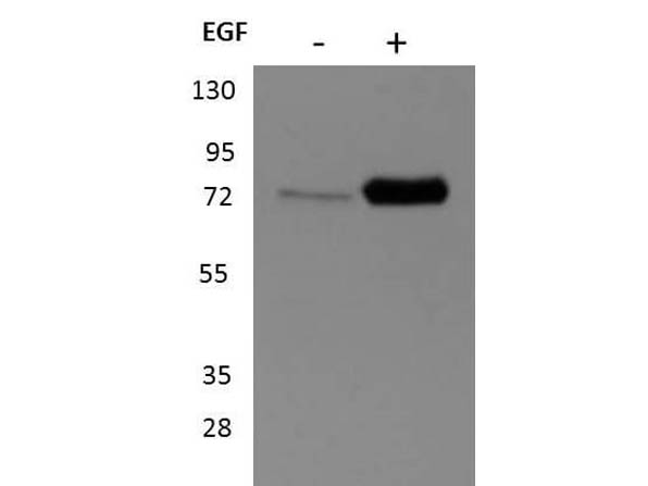
Western Blot of Rabbit anti-p90 RSK1 antibody. Lane 1: unstimulated HEK293T cell lysates. Lane 2: EGF stimulated HEK293T cell lysates. Load: 35 μg per lane.Primary antibody: p90 RSK1 antibody at 1:1000 for overnight at 4°C.Secondary antibody: peroxidase conjugated secondary antibody and ECL.Block: 5% BLOTTO overnight at 4°C.Predicted/Observed size: 90 kDa, ~90 kDa for p90 RSK1. Other band(s): none.
|
|
|
|
Western blot using Rockland's affinity purified anti-p90 RSK1 pS732 antibody shows detection of a band ~90 kDa in size corresponding to phosphorylated p90 RSK1 (arrowhead) in EGF stimulated (lane 2) HEK293T cell lysates prepared from cells grown in the absence of serum for 12 h. No staining is observed in similarly prepared lysates derived from unstimulated (control) cells (lane 1). After transfer, the membrane was blocked overnight followed by reaction with the primary antibody at a 1:1,000 dilution. Detection occurred using a peroxidase conjugated secondary antibody and ECL. Personal Communication. Kuldeep Patel, Loyola University Medical Center, Maywood, IL.
|
|
| 別品名 |
rabbit anti-p90 RSK1 pS732 antibody, rabbit anti-RSK1 pS732 antibody, Ribosomal S6 Kinase 1, RSK-1, S6K-alpha 1, 90 kDa Ribosomal Protein S6 Kinase 1, MAP kinase-activated protein kinase 1a, MAPK-activated protein kinase 1a, p90-RSK 1, p90S6K, MAPKAP kinase 1a, MAPKAPK-1a, Ribosomal S6 kinase 1, RSK 1, RPS6KA1, MAPKAPK1A
|
| 交差種 |
Human
|
| 適用 |
Western Blot
Enzyme Linked Immunosorbent Assay
|
| 免疫動物 |
Rabbit
|
| 抗原部位 |
C-terminus
|
| 標識物 |
Unlabeled
|
| 精製度 |
Affinity Purified
|
| 翻訳後修飾 |
リン酸化
|
| GENE ID |
6195
|
| Accession No.(Gene/Protein) |
NP_001006666.1, Q15418
|
| Gene Symbol |
RPS6KA1
|
| 参考文献 |
[Pub Med ID]24307699
|
| [注意事項] |
濃度はロットによって異なる可能性があります。メーカーDS及びCoAからご確認ください。
|
|
| メーカー |
品番 |
包装 |
|
RKL
|
600-401-B30S
|
25 UL
|
※表示価格について
| 当社在庫 |
なし
|
| 納期目安 |
約10日
|
| 保存温度 |
-20℃
|
|
※当社では商品情報の適切な管理に努めておりますが、表示される法規制情報は最新でない可能性があります。
また法規制情報の表示が無いものは、必ずしも法規制に非該当であることを示すものではありません。
商品のお届け前に最新の製品法規制情報をお求めの際はこちらへお問い合わせください。
|
※当社取り扱いの試薬・機器製品および受託サービス・創薬支援サービス(納品物、解析データ等)は、研究用としてのみ販売しております。
人や動物の医療用・臨床診断用・食品用としては、使用しないように、十分ご注意ください。
法規制欄に体外診断用医薬品と記載のものは除きます。
|
|
※リンク先での文献等のダウンロードに際しましては、掲載元の規約遵守をお願いします。
|
|
※CAS Registry Numbers have not been verified by CAS and may be inaccurate.
|
