|
※サムネイル画像をクリックすると拡大画像が表示されます。
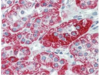
Immunohistochemistry using Rockland's anti-TRPC6 monoclonal antibody shows detection of TRPC6 in human adrenal (cortex) tissue (40X). The antibody was used a dilution to 2.5 μg/mL. The image shows strong staining with minimal background staining. Tissue was formalin fixed and paraffin embedded. No pre-treatment of sample was required. The image shows the localization of antibody as the precipitated red signal, with a hematoxylin purple nuclear counterstain. Personal communication, Andrew Elston, Lifespan Biosciences, Seattle, WA.
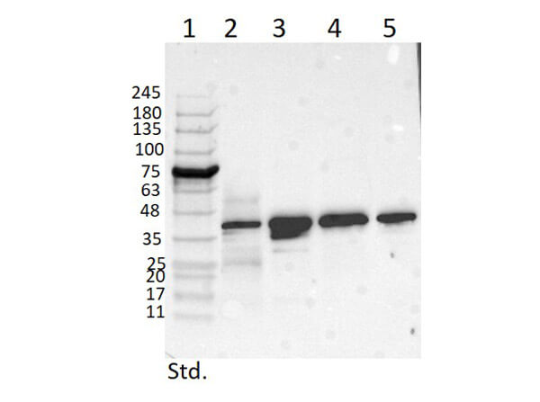
Western Blot of Mouse Anti-TRPC6 Antibody. Lane 1: Opal Prestained Molecular Weight Marker (p/n MB-210-0500). Lane 2: Mouse Pancreas Tissue Lysate (p/n W10-000-T023) [10μg]. Lane 3: MCF-7 Whole Cell Lysate (p/n W09-000-360) [10μg]. Lane 4: A431 Whole Cell Lysate (p/n W09-000-361) [10μg]. Lane 5: Jurkat Whole Cell Lysate (p/n W09-001-370) [10μg]. Primary Antibody: Anti-TRPC6 at 1μg/mL overnight at 2-8°C. Secondary Antibody: Rabbit Anti-Mouse IgG Peroxidase (p/n 610-403-C46) 1:40000 for 30mins at RT. Blocking Buffer: BlockOut Buffer (p/n MB-073) for 30mins at RT. Predicted MW: ~30kDa. Observed MW: ~40kDa. Exposure: 5sec.
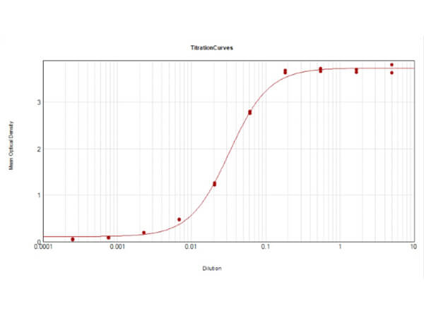
ELISA Results of Mouse Anti-TRPC6 Antibody. Each well was coated in duplicate with 0.1μg of conjugate. The working dilution is 1:31,000. The starting dilution of antibody was 5μg/ml and the X-axis represents the Log10 of a 3-fold dilution. This titration is a 4-parameter curve fit where the IC50 is defined as the titer of the antibody. Assay performed using HRP conjugation Stabilizer (p/n MB-076), Rabbit Anti-Mouse IgG HRP conjugated (p/n 610-403-C46) and TMB substrate (p/n TMBE-1000).
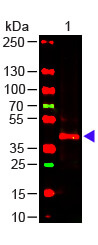
Western Blot of Mouse anti-TRPC6 Antibody. Lane 1: Mouse Kidney WCL (p/n W10-000-T017). Load: 10 μg per lane. Primary antibody: TRPC6 Antibody at 1:1000 for overnight at 4°C. Secondary antibody: donkey anti-mouse DyLight? 649 (p/n 610-743-002) at 1:20,000 for 30 min at RT. Block: MB-070 for 30 min at RT.
|
|
|
|
Immunohistochemistry using Rockland's anti-TRPC6 monoclonal antibody shows detection of TRPC6 in human adrenal (cortex) tissue (40X). The antibody was used a dilution to 2.5 μg/mL. The image shows strong staining with minimal background staining. Tissue was formalin fixed and paraffin embedded. No pre-treatment of sample was required. The image shows the localization of antibody as the precipitated red signal, with a hematoxylin purple nuclear counterstain. Personal communication, Andrew Elston, Lifespan Biosciences, Seattle, WA.
|
|
| 別品名 |
mouse anti-TRPC6 Antibody, TRPC 6, TRP6, short transient receptor potential channel 6 and transient receptor potential cation channel subfamily C member 6
|
| 交差種 |
Human
Mouse
|
| 適用 |
Western Blot
Enzyme Linked Immunosorbent Assay
Immunohistochemistry
|
| 免疫動物 |
Mouse
|
| クローン |
3F2.H10.F2
|
| 抗体クラス |
IgG1κ
|
| 抗原部位 |
C-terminus
|
| 標識物 |
Unlabeled
|
| 精製度 |
Ig fraction - Protein A
|
| GENE ID |
7225
|
| Accession No.(Gene/Protein) |
5730102, Q9Y210
|
| Gene Symbol |
TRPC6
|
| 参考文献 |
[Pub Med ID]27874267
|
| [注意事項] |
濃度はロットによって異なる可能性があります。メーカーDS及びCoAからご確認ください。
|
|
| メーカー |
品番 |
包装 |
|
RKL
|
200-301-B59S
|
25 UL
|
※表示価格について
| 当社在庫 |
なし
|
| 納期目安 |
約10日
|
| 保存温度 |
-20℃
|
|
※当社では商品情報の適切な管理に努めておりますが、表示される法規制情報は最新でない可能性があります。
また法規制情報の表示が無いものは、必ずしも法規制に非該当であることを示すものではありません。
商品のお届け前に最新の製品法規制情報をお求めの際はこちらへお問い合わせください。
|
※当社取り扱いの試薬・機器製品および受託サービス・創薬支援サービス(納品物、解析データ等)は、研究用としてのみ販売しております。
人や動物の医療用・臨床診断用・食品用としては、使用しないように、十分ご注意ください。
法規制欄に体外診断用医薬品と記載のものは除きます。
|
|
※リンク先での文献等のダウンロードに際しましては、掲載元の規約遵守をお願いします。
|
|
※CAS Registry Numbers have not been verified by CAS and may be inaccurate.
|
